Microscopy
90 images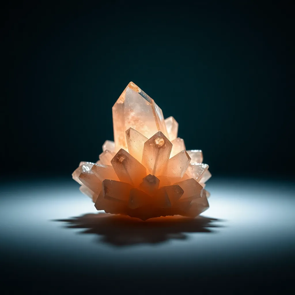
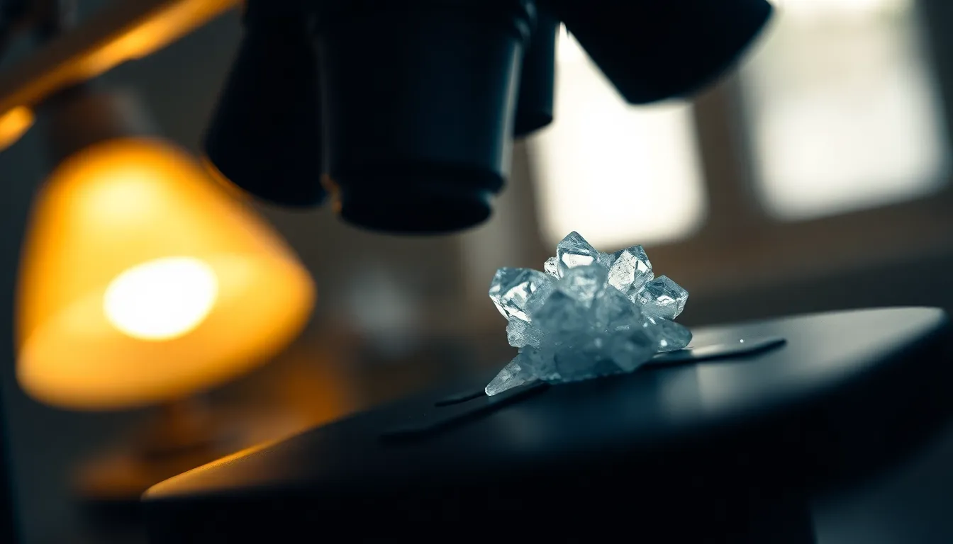
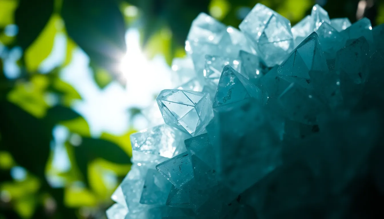
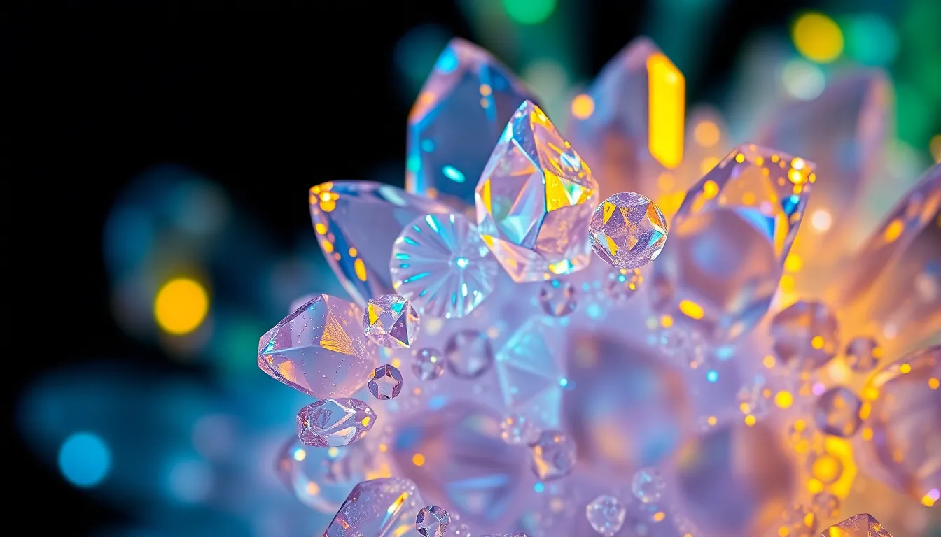
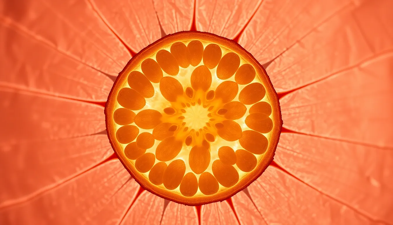
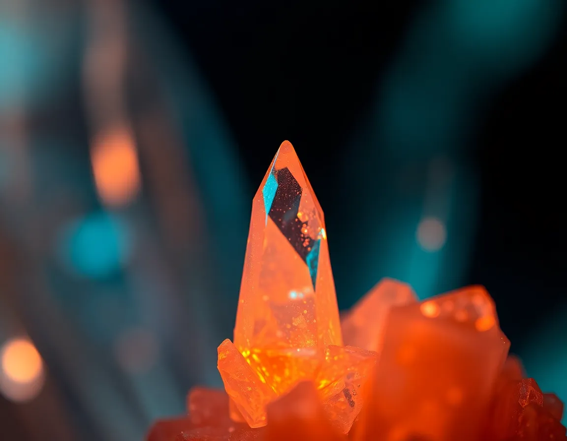
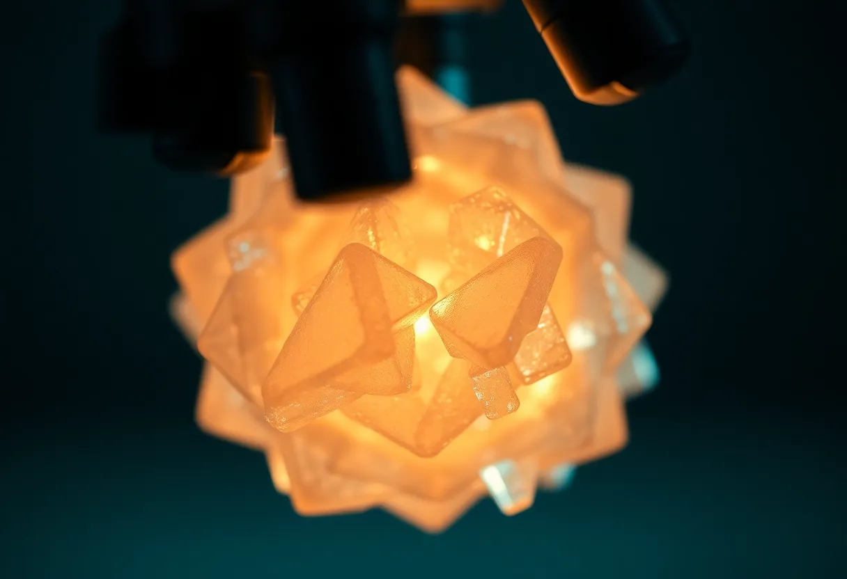
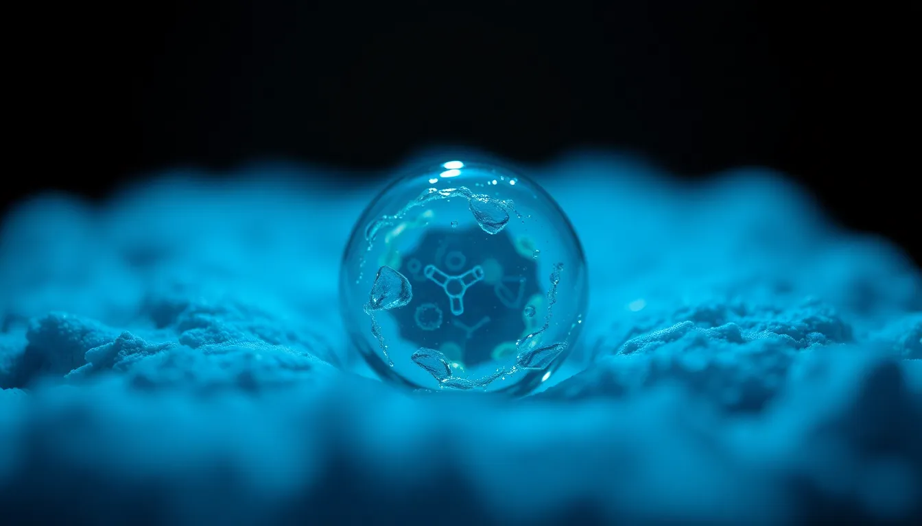
shot on Leica Q3, 28mm f/1.7 Summilux, featuring an intense spotlight on a uniquely structured crystal with soft fill light providing subtle highlights, shallow depth of field at f/1.7 creating an ethereal bokeh, cinematic teal and orange color grading enhancing the contrast, centered symmetrical composition focusing on the crystal structure's facets and texture details, with a dark, smooth background to emphasize the subject.
1:1
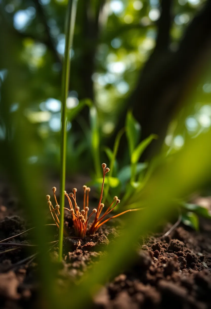
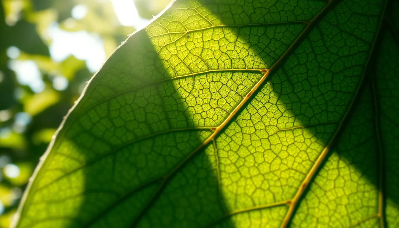
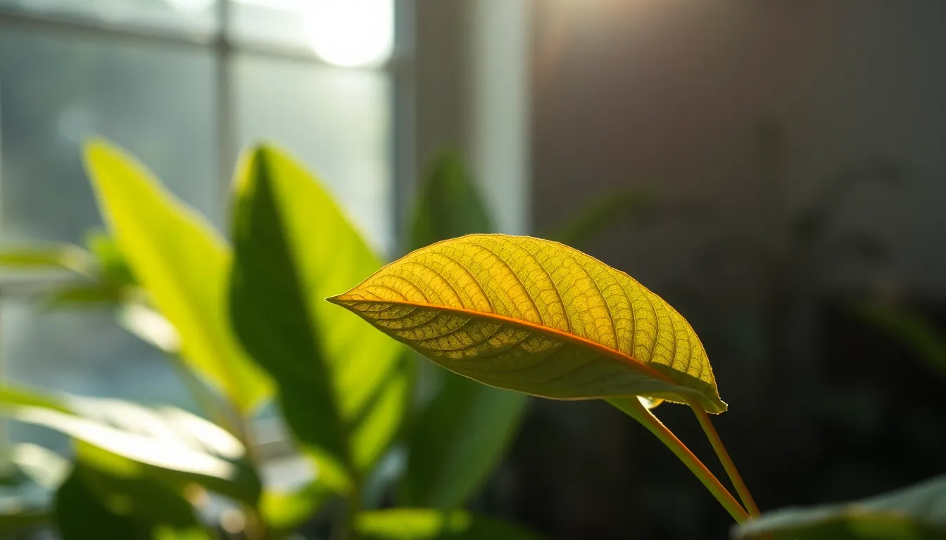
Fujifilm GFX 100S, 110mm f/2 macro, captured in diffused daylight filtering through tree canopy creating dappled light effects, shallow depth of field at f/2 with creamy bokeh surrounding the specimen, natural muted tones with rich greens and browns, composition utilizing leading lines formed by plant material guiding the viewer's eye toward a microscopic view of soil microbes with visible texture details.
2:3
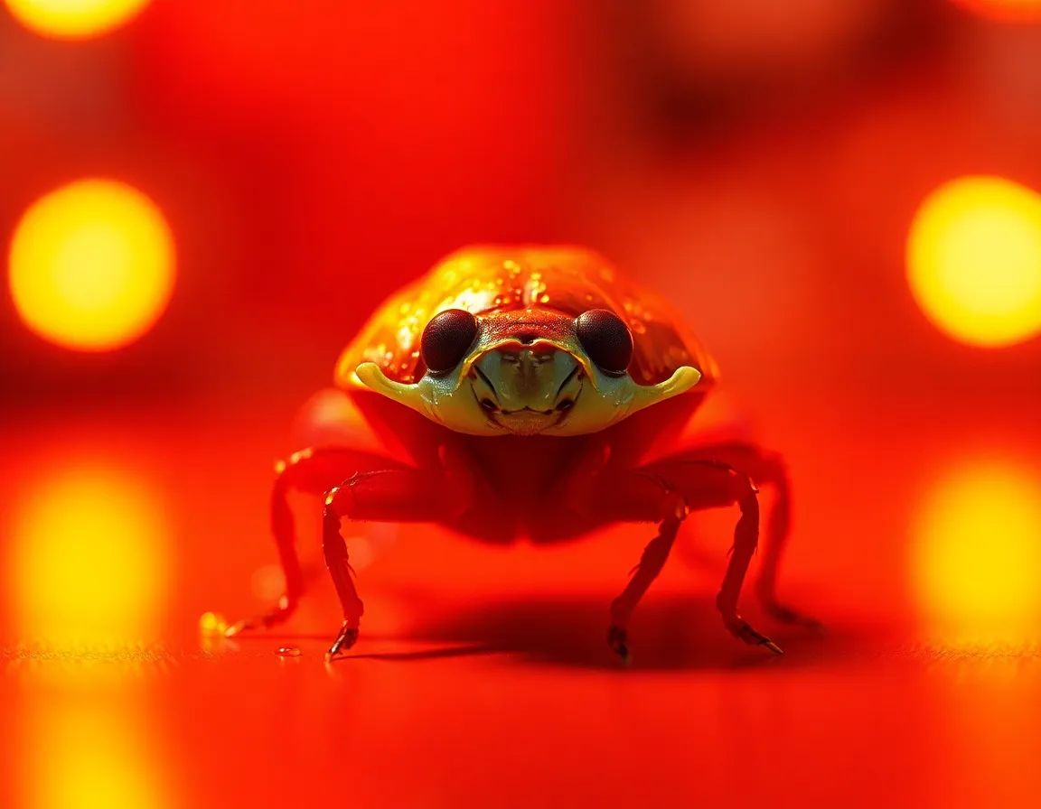
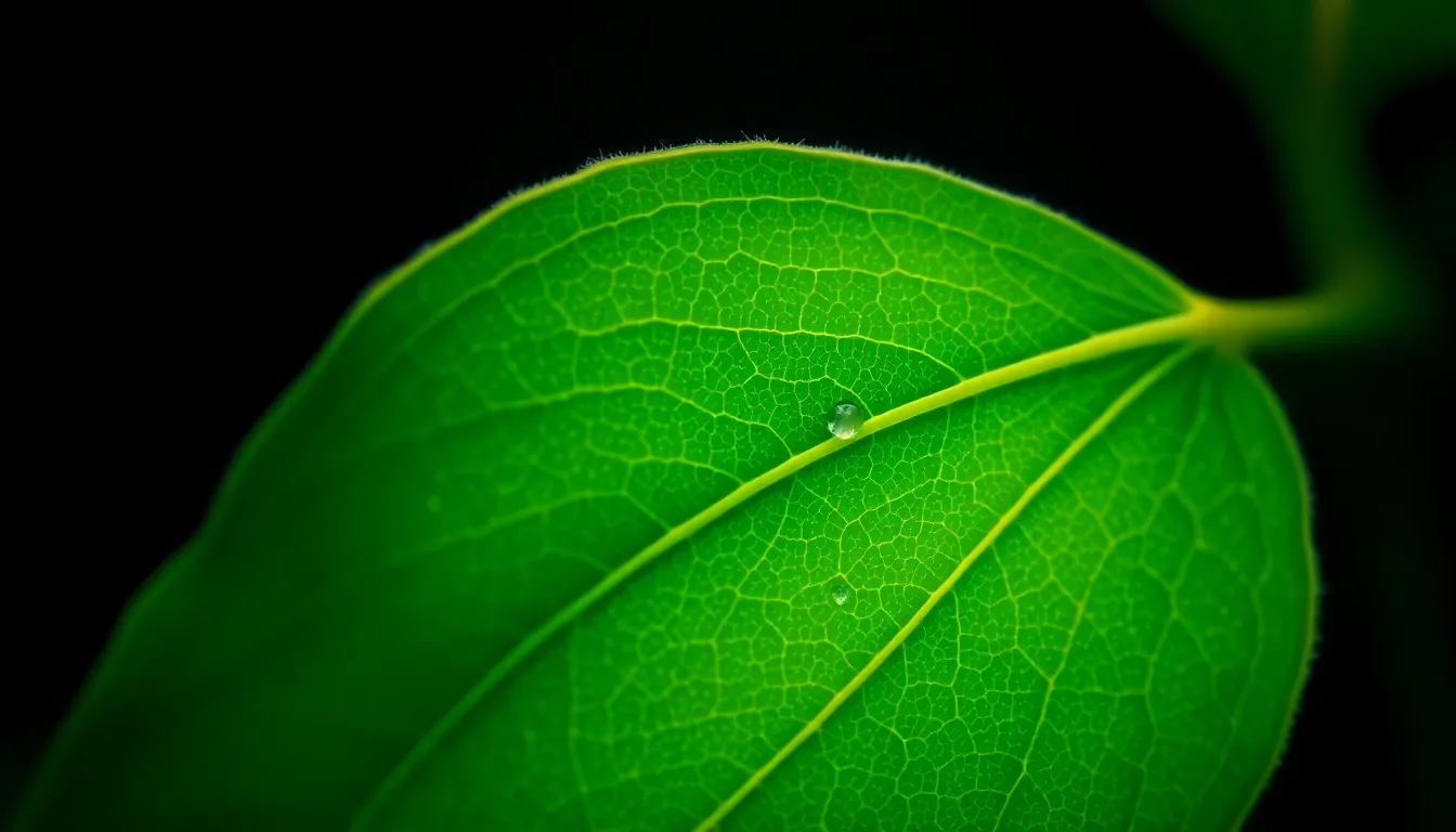
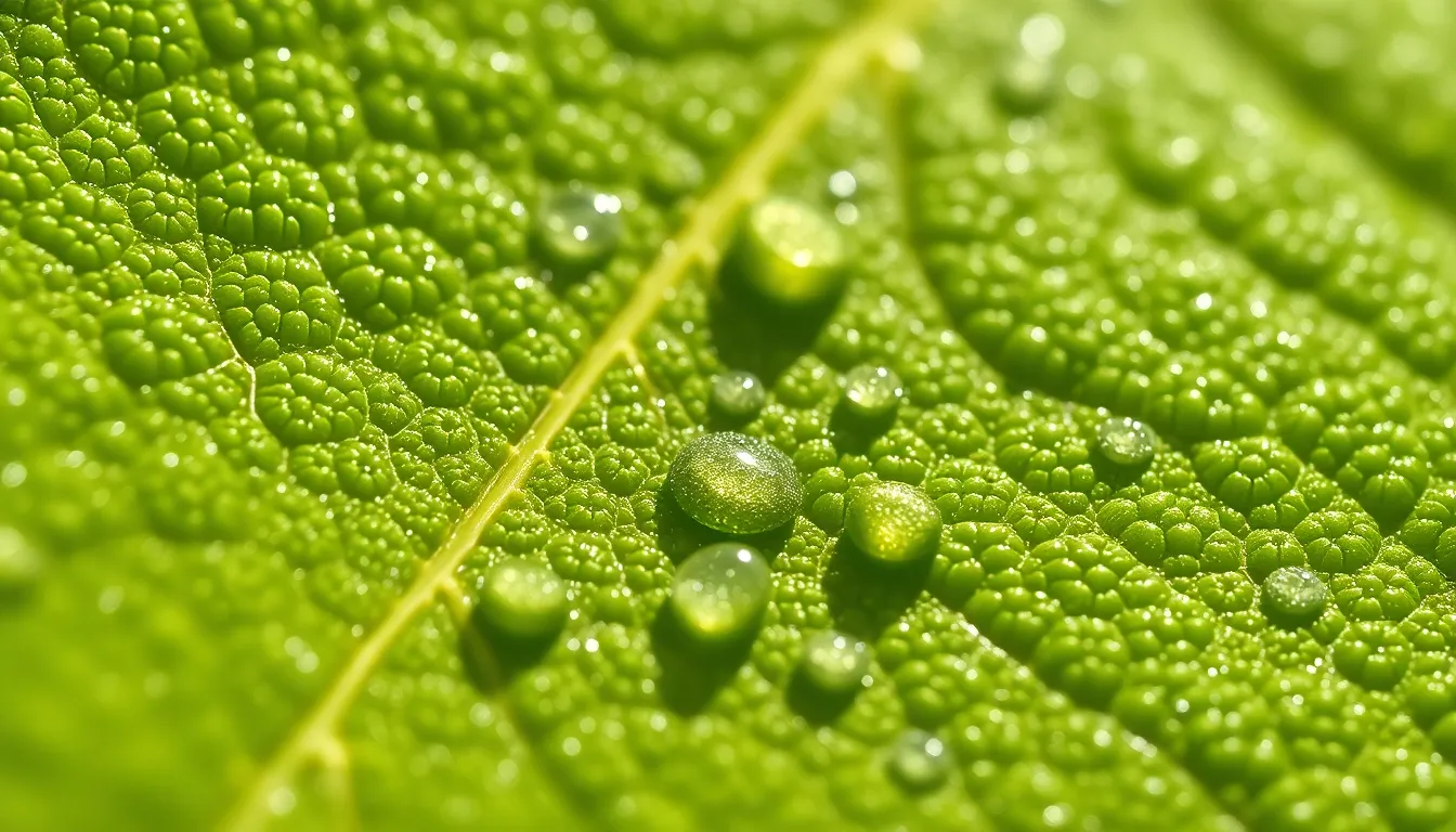
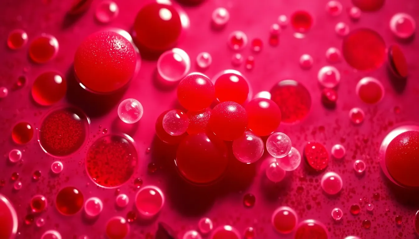
Hasselblad X2D 100C, 90mm f/2.5 medium format, lit by a practical warm tungsten lamp casting directional light, selective focus on a vibrant organism with the background softening into a painterly bokeh, Kodachrome-inspired saturation boosting colors to a lively palette, centered symmetrical composition highlighting the subject's symmetry and details, with visible water droplets on the specimen enhancing the vividness.
4:3
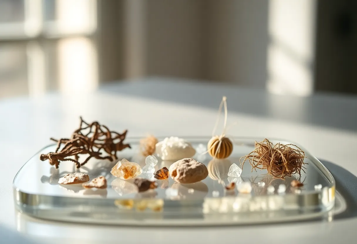
captured with Canon EOS R5, 24-70mm f/2.8L at 50mm, soft natural daylight diffusing through a window creating gentle highlights, hyperfocal distance ensuring everything from foreground to infinity is tack-sharp at f/8, natural muted tones with desaturated earth colors, composition focusing on a prepared slide with various samples arranged artistically, showcasing different textures on the glass surface, including tiny crystals and fibers.
3:2
shot on Nikon Z9, 70-200mm f/2.8 telephoto at 135mm, illuminated by bright LED ring light enhancing specimen details, shallow depth of field at f/2.8 with a creamy bokeh background, vibrant Fujifilm Velvia-inspired color palette emphasizing deep blues and rich greens, composition through a close-up on a single cell structure showcasing intricate patterns and textures, with a glass slide reflecting light subtly beneath the specimen.
16:9
Fujifilm GFX 100S, 110mm f/2 macro, studio lighting with three-point setup featuring a key softbox creating even light across the specimen, hyperfocal distance producing a sharp image from foreground to background, Fujifilm Velvia-inspired saturated colors with vivid reds and greens, rule of thirds composition with the specimen strategically placed at an intersection point, highlighting intricate textures and details on the slide.
16:9
Hasselblad X2D 100C, 90mm f/2.5 medium format, dappled sunlight filtering through tree canopy creating speckled light on the transparent slide, shallow depth of field at f/2.5 focusing sharply on the central specimen while blurring surroundings, natural muted tones with desaturated colors enhancing the organic feel, foreground framing with leaves surrounding the slide for a cohesive natural composition, and visible texture of the glass reflecting light.
16:9

captured with Canon EOS R5, 24-70mm f/2.8L at 50mm, golden hour backlighting with warm rim light highlighting the edges of the prepared specimen, hyperfocal distance producing tack-sharp focus throughout the scene, cinematic teal and orange color grading creating a dramatic contrast, leading lines guiding the viewer's eye toward the specimen, with detailed textures of the glass and sample visible.
16:9
shot on Sony A7R V with 85mm f/1.4 GM lens, warm tungsten desk lamp casting directional light onto the prepared slide, selective focus on the vibrant specimen, background gradually melting into soft circular bokeh, Kodak Portra 400 color palette enhancing warm tones and creamy highlights, rule of thirds composition with the specimen positioned at the left power point, showcasing the smooth glass slide with its reflections.
16:9
captured with Nikon Z9, 70-200mm f/2.8 telephoto at 100mm, overcast diffused daylight illuminating the specimen slide from an overhead light source, shallow depth of field at f/2.8 with a sharp focus on the specimen and a softly blurred background, natural muted tones with desaturated earth colors, centered symmetrical composition highlighting the glass slide, and visible texture of the specimen with intricate details on cellular structures.
16:9
shot on Leica Q3, 28mm f/1.7 Summilux, featuring warm tungsten desk lamp casting directional pool of light on a glass slide with human cells. Shallow depth of field at f/1.7 creates an ethereal bokeh effect with a softly blurred background. The color palette features warm tones of amber and golden highlights, evoking a cozy, intimate atmosphere. The composition follows the rule of thirds, placing the slide in a power position to draw the eye, while the textures of the glass and the cells' surfaces come alive under the warm light.
9:16
captured with Hasselblad X2D 100C, 90mm f/2.5 medium format, using natural, overcast diffused daylight to create soft lighting. Hyperfocal distance at f/8 ensures all details of a micro-fossil specimen are sharp and clear. The color palette reveals subtle earth tones, with beige and muted browns enhancing the organic feel of the fossil. The composition follows leading lines that draw the viewer's gaze along the natural formations of the fossil, emphasizing its intricate textures.
3:4
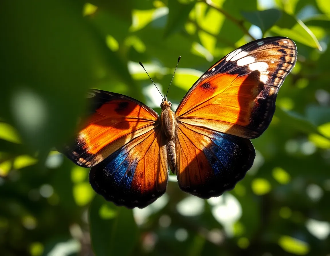
shot on Fujifilm GFX 100S, 110mm f/2 macro, illuminated by dappled sunlight filtering through leaves, creating a natural and soft environment. Shallow depth of field at f/2 enhances the intricate patterns of butterfly wings viewed under a microscope. The rich color palette features vibrant blues, oranges, and subtle browns, emphasizing the beauty and complexity of the wing structure. The composition is centered, allowing the viewer to delve into the captivating details that define this natural specimen.
4:3
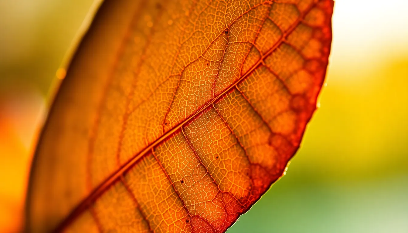
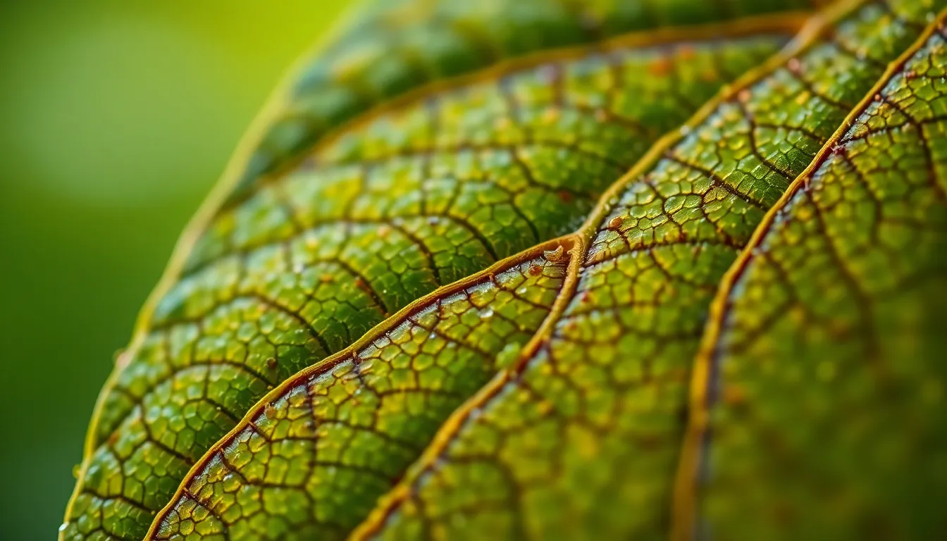
captured with Canon EOS R5, 24-70mm f/2.8L at 50mm, illuminated by a three-point studio setup with key softbox creating focused light and subtle fill. Hyperfocal distance at f/8 ensures everything is sharply defined, showcasing a detailed cross-section of a leaf under optical microscopy. The natural muted tones of the leaf are highlighted with deep greens and browns against a contrasting black background. The composition adheres to the rule of thirds with the leaf positioned to draw attention, creating a sense of depth.
3:2
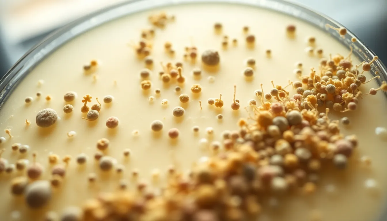
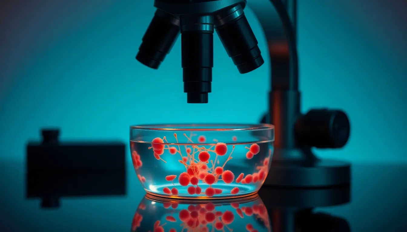
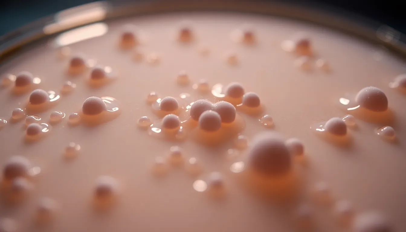
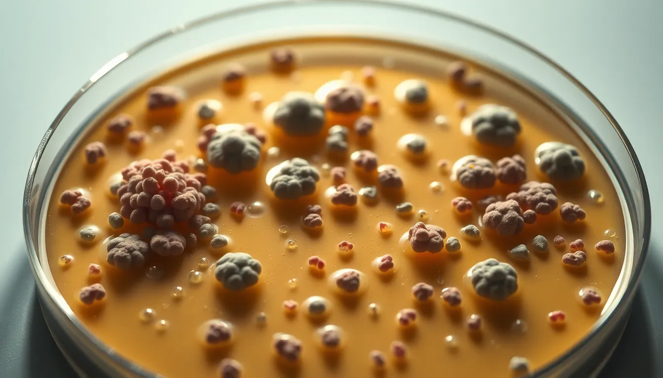
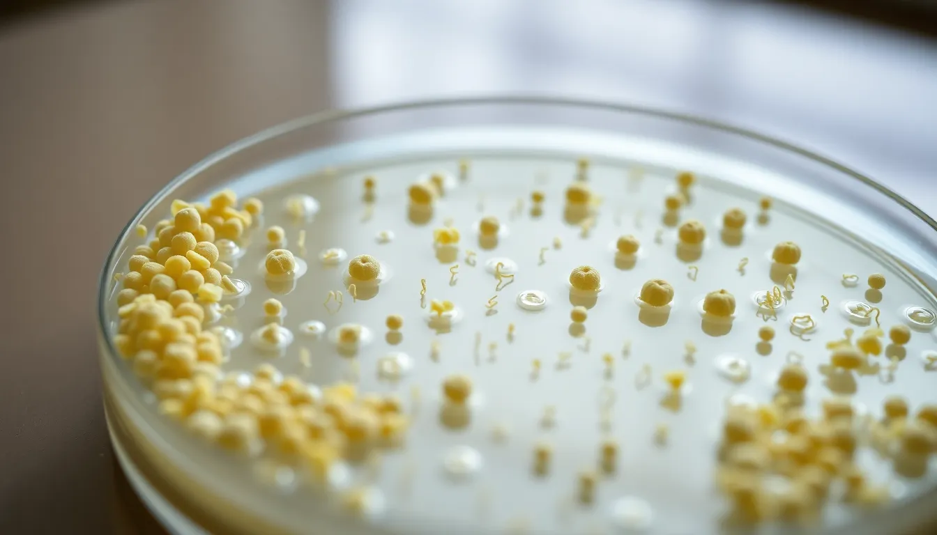
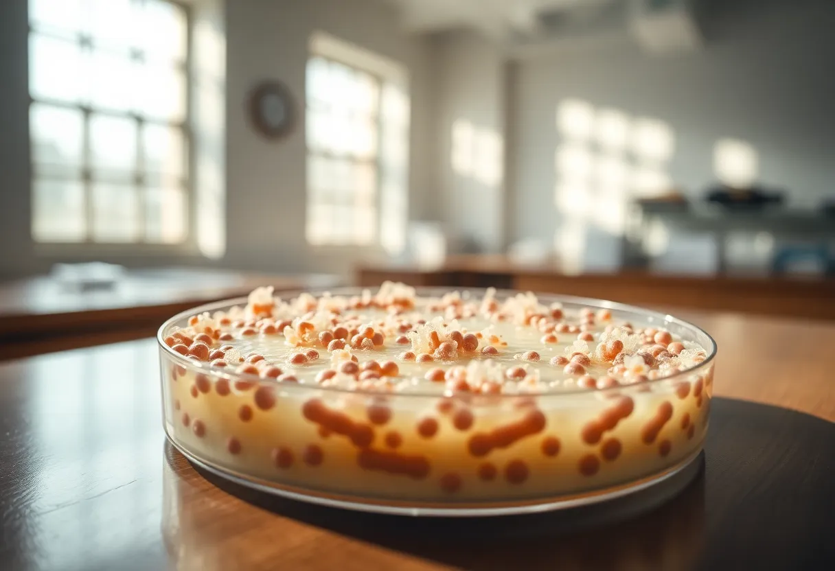
shot on Nikon Z9, 70-200mm f/2.8 telephoto at 135mm, illuminated by soft, diffused daylight filtering through a microscope light source. Shallow depth of field at f/2.8 highlights the intricate details of a petri dish filled with colorful bacterial cultures. The vivid color palette reflects saturated greens and yellows, emphasizing the biological diversity. Composition captures the dish slightly off-center, creating a dynamic visual flow. The smooth glass of the dish contrasts with the textured surface of the microscope stage, lending a scientific elegance to the image.
16:9
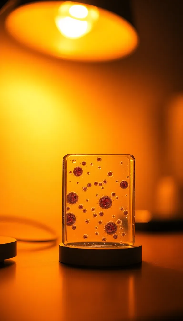
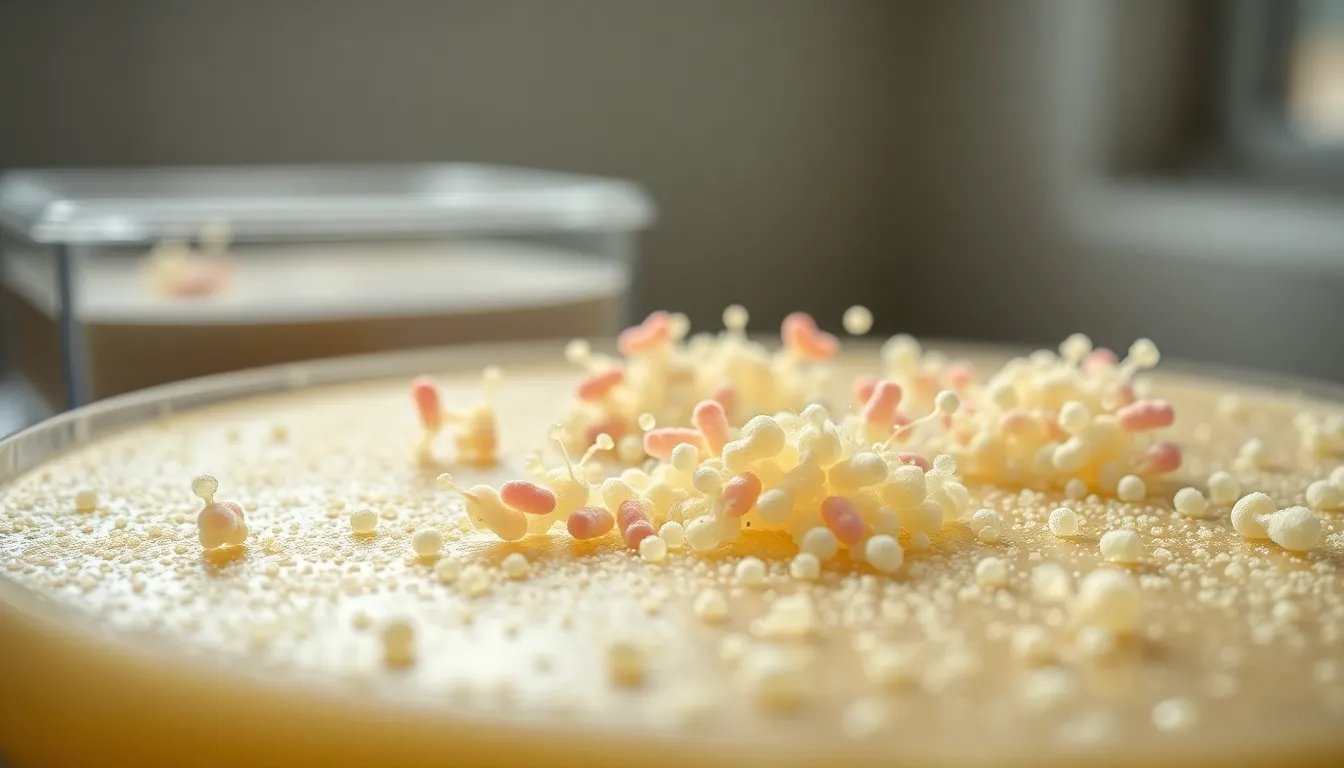
Photographed with Fujifilm GFX 100S, 110mm f/2 macro, this image displays a close-up view of bacteria colonies growing on agar gel. The lighting is provided by a warm tungsten desk lamp, creating a dramatic effect that highlights the bacteria's shapes and colors. The depth of field is shallow at f/2, producing a luxurious bokeh in the background that isolates the bacteria. The color grading is cinematic with rich contrast, enhancing the vivid colors of the colonies. The composition is centered, with the bacteria colonies standing out sharply against the smooth agar surface, showcasing their intricate details.
16:9
Created with Hasselblad X2D 100C, 90mm f/2.5 medium format, this image presents intricate patterns of a leaf's cellular structure viewed under a microscope. The lighting is natural, with dappled sunlight filtering through surrounding leaves, creating a soft, ethereal glow. The image utilizes hyperfocal distance, keeping everything from the foreground to the leaf's intricate cells in sharp focus at f/8. The color palette consists of natural muted tones, emphasizing the greens and browns of the leaf. The composition follows the rule of thirds, placing the leaf's veins along the lines, guiding the viewer's eye through the detailed structures.
16:9
Captured using Nikon Z9, 70-200mm f/2.8 telephoto at 135mm, this image portrays a microbe on a petri dish, showcasing its unique shapes and textures. The lighting is provided by a warm tungsten desk lamp, casting a directional pool of light that accentuates the microbe's surface details. The depth of field is shallow at f/2.8, with a soft bokeh background that emphasizes the subject. The color science follows a natural muted palette with desaturated earth colors, creating an organic feel to the image. The composition employs leading lines, directing the viewer's attention toward the microbe, positioned prominently in the center.
16:9

Shot on Sony A7R V with 85mm f/1.4 GM lens, this image features a detailed view of crystalline structures under polarized light. The lighting is a mix of vibrant colors generated by refraction, creating a spectrum of hues across the crystals' surfaces. The image utilizes a shallow depth of field at f/1.4, which beautifully blurs the background while keeping the crystals sharply in focus. A Kodak Portra 400 color palette adds warmth to the overall image, enhancing the natural colors of the crystal formations. The composition is centered, providing symmetry in the arrangement of the crystals, which appear to shimmer with intricate patterns and textures.
16:9
Captured with Canon EOS R5, 24-70mm f/2.8L at 70mm, the image showcases a vibrant cellular structure under a microscope. Soft, diffused daylight filters through a laboratory window, providing natural illumination that highlights the intricate details of the cells. The image features a shallow depth of field at f/2.8, creating a creamy bokeh effect in the background, emphasizing the cellular textures. The color palette is rich with deep blues and vivid greens, inspired by Fujifilm Velvia. The composition follows the rule of thirds, placing a single, detailed cell in the right power point, drawing the viewer's gaze. Textures of the microscopic sample appear almost tangible, with visible boundaries and contours illuminated by the light.
16:9
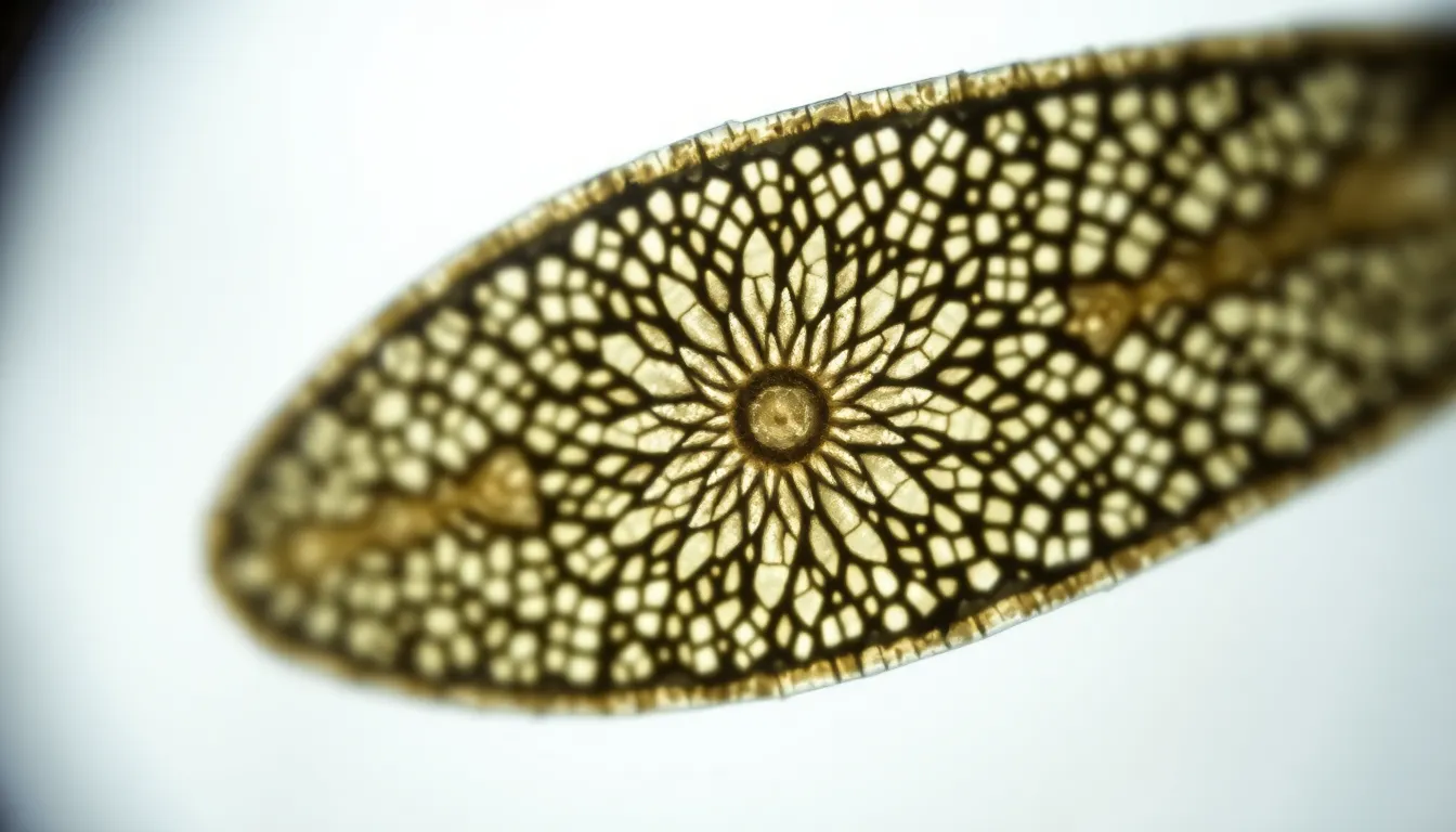
Detailed view of a diatom under a microscope, showcasing its intricate silica structure. Shot with Hasselblad X2D 100C, 90mm f/2.5 medium format lens. Natural light from a nearby window provides soft illumination, enhancing the delicate detail of the diatom's pattern. Depth of field at f/5 creates a sharp focus on the diatom while blurring the background glass slide. The composition uses centered symmetry to draw attention to the stunning natural design. The texture of the diatom's surface is highlighted, exhibiting its unique crystalline structure.
16:9
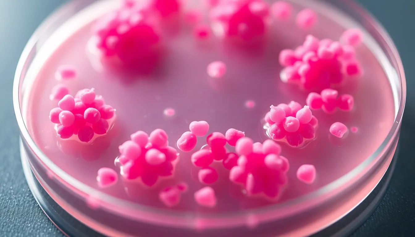
Microscopic view of bacteria colonies on an agar plate. Captured with Leica Q3, 28mm f/1.7 Summilux lens. The scene is illuminated with soft overhead studio lighting, enhancing the contrast of the vibrant pink and white bacterial colonies against the clear agar surface. Shallow depth of field at f/2.8 isolates the colonies, creating a creamy bokeh that emphasizes their unique shapes. The composition follows the rule of thirds, placing the most prominent colony at a power point. Textural details of the agar surface are visible, adding depth.
16:9
Detailed view of a crystal structure under polarized light microscopy. Shot on Fujifilm GFX 100S, 110mm f/2 macro lens. Bright and colorful patterns emerge from the polarized light, creating stunning geometric shapes. Selective focus on the central crystal formation, while the background blurs into a soft, painterly bokeh. The color palette features vibrant blues, purples, and yellows, reminiscent of a vibrant gemstone. The composition utilizes symmetry, drawing attention to the striking design of the crystal's facets.
16:9
Close-up view of a leaf's surface revealing trichomes and stomata under a microscope. Captured with Canon EOS R5, 24-70mm f/2.8L at 50mm. Natural diffused daylight floods the scene, creating soft highlights and shadows across the leaf's textured surface. Hyperfocal distance — everything from the foreground trichomes to the background stomata is tack-sharp at f/8. The composition follows the rule of thirds, focusing on the intricate details of the leaf. Visible texture of the leaf's surface with clear water droplets enhancing the lush green color.
16:9
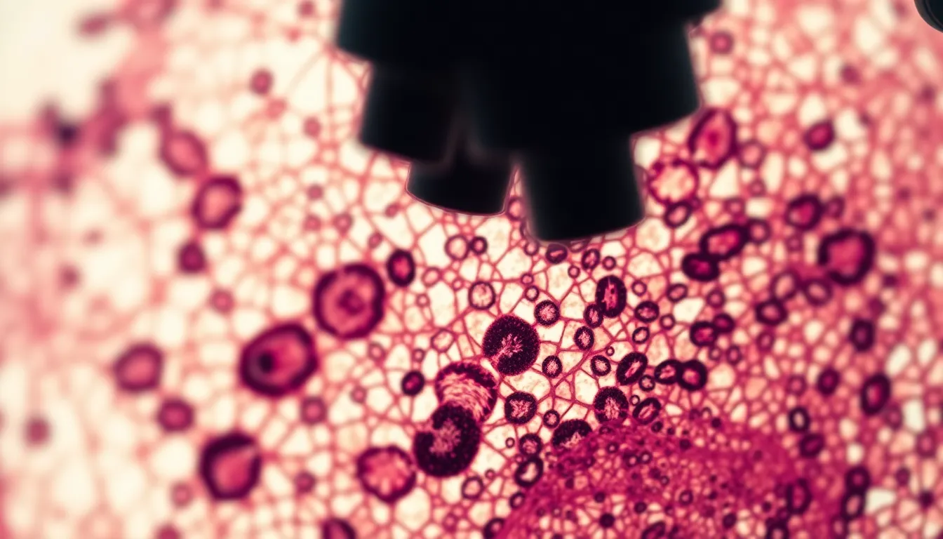
Microscopic view of a human blood smear showcasing various blood cells. Shot on Nikon Z9, 70-200mm f/2.8 telephoto at 135mm. Backlit with a bright field microscope illumination, creating high contrast between cells and background. Shallow depth of field at f/2.8, with erythrocytes displaying rich red tones and leukocytes appearing soft white against a dark backdrop. Composition centered on a cluster of cells with leading lines created by the slide's structure. Textures of the slide surface visible alongside the smooth gel of the blood specimen.
16:9
captured with Leica Q3, 28mm f/1.7 Summilux, lit with dramatic back-lighting from a warm desk lamp casting long shadows, selective focus on a detailed microscope slide showcasing a crystalline substance, natural muted tones with deep blues and grays enhancing the clarity of the crystals, leading lines formed by the edges of the microscope directing the eye towards the focal point of the slide, capturing the intricate surface texture of the crystalline material.
16:9
shot on Hasselblad X2D 100C, 90mm f/2.5 medium format, illuminated with a softbox providing soft, even light, tilt-shift miniature effect with a narrow band of focus on a cluster of microorganisms, natural muted tones with desaturated pastels creating a dreamy atmosphere, centered symmetrical composition with the microorganisms arranged artfully within the frame, the textured surface of the microscope stage enhancing the overall depth.
16:9
captured with Canon EOS R5, 24-70mm f/2.8L at 35mm, illuminated by soft ambient daylight coming from a large window creating gentle shadows, shallow depth of field at f/2.8 focusing on a colorful cross-section of a leaf, natural muted tones with desaturated greens and yellows, rule of thirds composition positioning the leaf slice at the right power point while the background softly blurs into an organic green bokeh of the surrounding plant life.
16:9
captured with Fujifilm GFX 100S, 110mm f/2 macro, dramatic overhead studio lighting with a single softbox creating crisp shadows, hyperfocal distance ensuring all details from the petri dish to the microscope are sharp at f/8, cinematic teal and orange color grading enhancing visual interest, leading lines created by the microscope’s base directing the eye to vibrant bacterial cultures in the dish, the smooth glass surface reflecting the surrounding colors.
16:9
shot on Nikon Z9, 70-200mm f/2.8 telephoto at 135mm, illuminated under bright LED microscopy lights providing high contrast, shallow depth of field at f/2.8 featuring a creamy bokeh background, natural muted tones with desaturated earth colors, centered symmetrical composition highlighting a vibrant stained tissue sample with visible cellular structures, the glossy surface of the slide reflecting light and enhancing details.
16:9
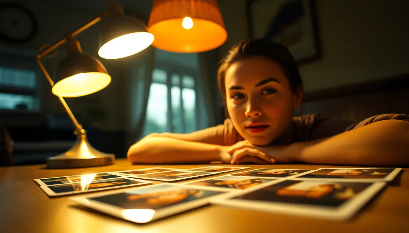
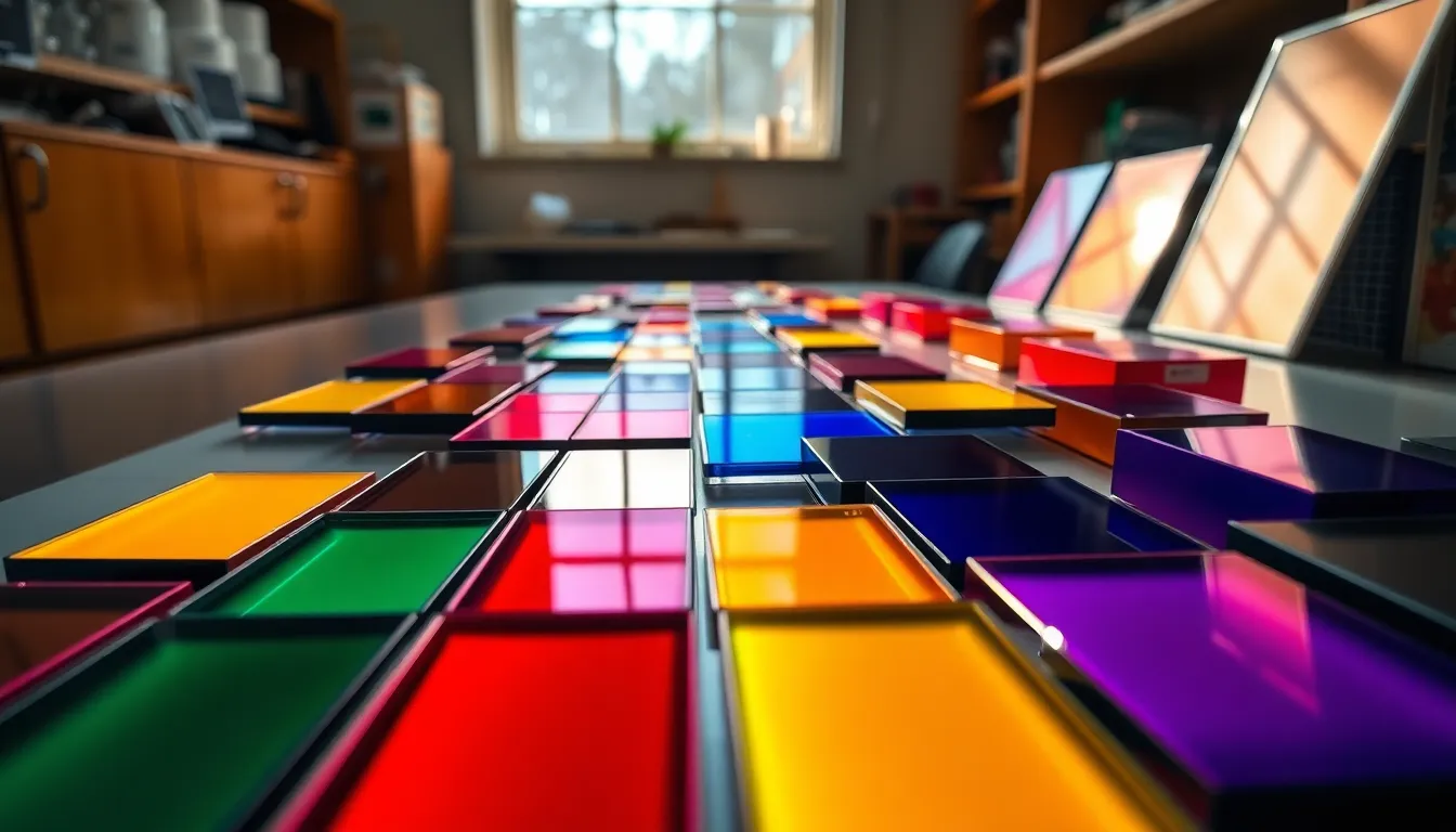
captured with Leica Q3, 28mm f/1.7 Summilux, illuminated by a warm practical lamp providing a cozy pool of light, shallow depth of field at f/1.7 with creamy bokeh in the background, Kodak Portra 400 color palette emphasizing warm skin tones and creamy highlights, Dutch angle composition creating dynamic tension while showcasing various slides laid out on a wooden table.
16:9
shot on Hasselblad X2D 100C, 90mm f/2.5 medium format, illuminated by overcast diffused daylight through large windows, hyperfocal distance achieving crisp detail from foreground to background, natural muted tones with soft earth colors, centered symmetrical composition with a petri dish showcasing visible crystal formations, detailed surface textures reflecting light subtly.
16:9
captured with Canon EOS R5, 24-70mm f/2.8L at 35mm, illuminated by under-table warm tungsten lighting creating dramatic shadows, shallow depth of field at f/2.8 with a creamy bokeh backdrop of laboratory equipment, cinematic teal and orange color grading enhancing the mood, rule of thirds composition with the specimen jar positioned at the left power point, showcasing detailed glass texture and liquid movement.
16:9
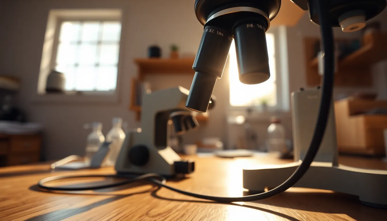
shot on Fujifilm GFX 100S, 110mm f/2 macro lens, under diffused natural daylight streaming through a window, hyperfocal distance ensuring everything from foreground to infinity is tack-sharp, natural muted tones with desaturated earth colors, composition using leading lines of microscope cables drawing the eye towards the eyepiece, capturing the soothing texture of wood on the laboratory desk.
16:9
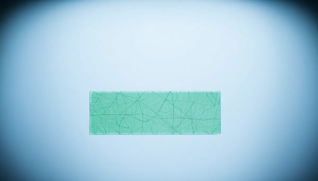
captured with Nikon Z9, 70-200mm f/2.8 telephoto at 135mm, illuminated by a bright LED ring light providing even illumination across the specimen, shallow depth of field at f/2.8 creating a soft bokeh background, pharmaceutical green and sterile white color palette, centered symmetrical composition, featuring textured glass slides with visible scratches reflecting light.
16:9
An intricate view of a crystalline structure, captured with Leica Q3, 28mm f/1.7 Summilux lens. The lighting is a mix of dappled sunlight filtering through leaves, creating a playful pattern of highlights and shadows on the crystal. The depth of field is shallow at f/1.7, providing a dreamy bokeh against the sharp foreground. The color palette features cool blues, greens, and hints of sparkling white from the crystals. The composition emphasizes the crystal’s geometric shapes and patterns, utilizing a Dutch angle for dynamic tension. The texture is vivid, reflecting facets and surfaces that catch light beautifully.
16:9
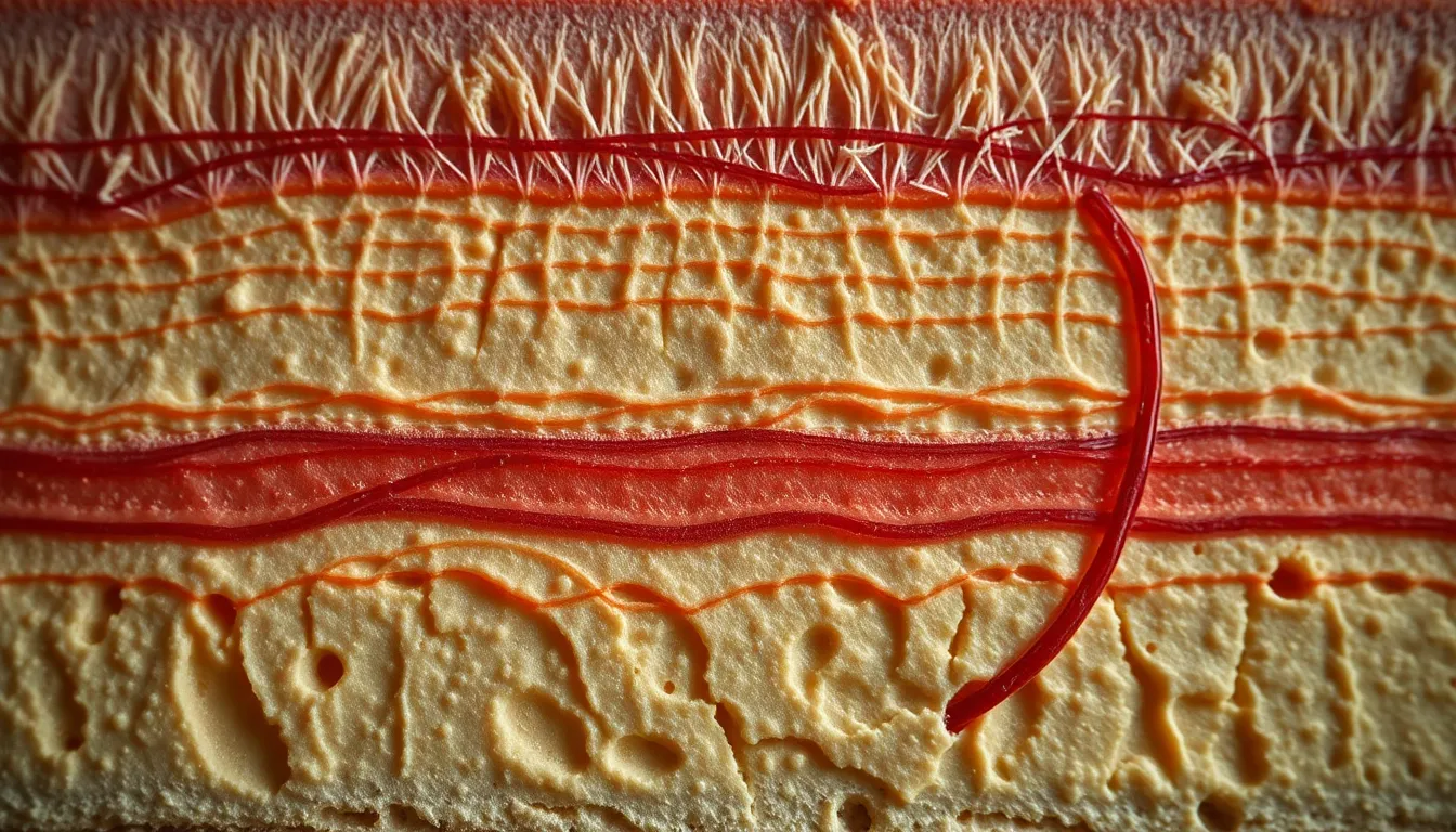
A detailed view of a cross-section of human skin, shot on Hasselblad X2D 100C, 90mm f/2.5 medium format lens. The lighting features a practical setup, utilizing warm tungsten desk lamps to create a gentle glow that adds depth to the texture. The shot employs hyperfocal distance at f/8, ensuring sharp focus throughout the entire frame. The color palette is natural and muted, with creamy skin tones and subtle reds showing blood vessels. Compositionally, the image utilizes leading lines to guide the viewer’s eye through the layers of skin. The texture is portrayed with remarkable detail, highlighting pores, hair follicles, and the unique patterns of skin cells.
16:9
A dynamic view of a plant cell, captured using Fujifilm GFX 100S, 110mm f/2 macro lens. The lighting is provided by a three-point studio setup, with key light highlighting the cell structure, edge light creating separation, and subtle fill light enhancing textures. The depth of field is shallow at f/2, allowing the nucleus and chloroplasts to stand out sharply against a softly blurred background. The color palette features natural greens and earthy browns, invoking the essence of a botanical subject. The composition follows a centered symmetrical layout, drawing the viewer directly to the cell's intricate features. Textures are finely detailed, showcasing the cell walls and organelles in stunning clarity.
16:9
An isolated view of bacterial colonies grown on agar media, shot on Canon EOS R5, 24-70mm f/2.8L at 50mm. The scene is lit with natural overcast daylight filtering through a window, providing soft, diffused illumination. A selective focus on the colonies at f/4 blurs the background slightly, directing attention to the unique shapes and colors of the bacteria. The color palette is natural with earthy tones and splashes of pastel hues from the colonies. Compositionally, the image adheres to the rule of thirds, positioning the most vibrant colonies at one of the power points. The details reveal the texture of the agar with fine granules, demonstrating the growth environment.
16:9
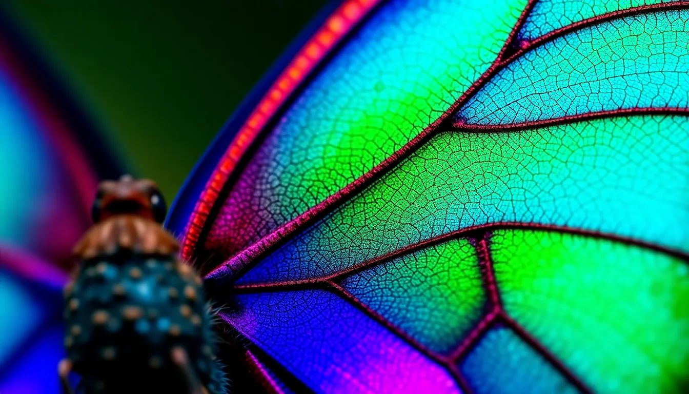
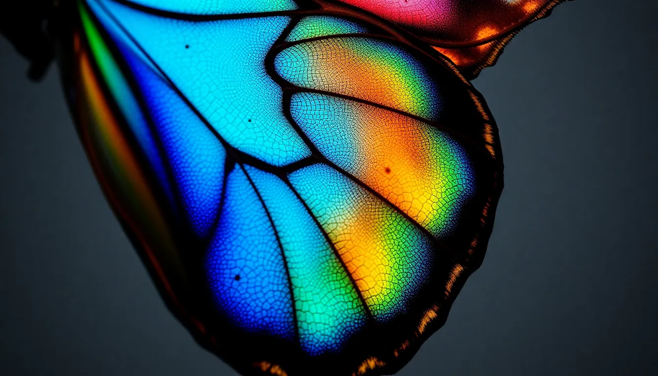
A photomicrograph of a vibrant butterfly wing, captured with a Nikon Z9, 70-200mm f/2.8 telephoto at 200mm. The lighting is provided by a bright LED ring light, creating an even illumination that enhances the color and detail of the scales. The image features a shallow depth of field at f/2.8, allowing for a soft bokeh effect in the background. The color palette is rich and saturated, showcasing vivid blues, greens, and iridescent purples. Compositionally, the subject is slightly off-center, allowing for negative space to enhance the delicate details of the wing's patterns. The texture is emphasized through the fine detail of the scale surface, reflecting light beautifully.
16:9
Shot on Hasselblad X2D 100C, 90mm f/2.5 medium format, utilizing overcast daylight through a large window for soft, diffused lighting. The image presents a close-up view of bacteria cultures on an agar plate, showcasing intricate formations and textures. The depth of field is shallow at f/2.5, bringing the focus to the most vibrant bacterial colonies while blurring the edges. The color palette features muted earth tones with hints of vibrant greens and yellows. The composition uses a diagonal angle, creating dynamic tension with leading lines formed by the bacterial growth patterns.
16:9
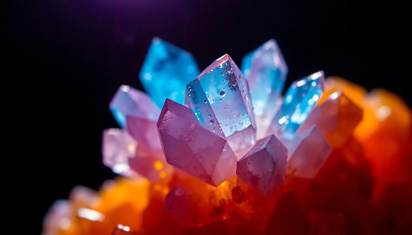
Captured with Fujifilm GFX 100S, 110mm f/2 macro, showcasing a stunning crystalline structure under polarized light, creating vivid colors. The lighting is dramatic, providing stark contrasts that reveal the intricate details of the crystal formation. The image employs a shallow depth of field at f/2, emphasizing the foreground crystal and blurring out the background. The color palette is inspired by Fujifilm Velvia, filled with saturated colors of blues, purples, and fiery oranges. The composition is centered, drawing attention to the crystal's facets and textures, which resemble a carefully crafted sculpture.
16:9
Photographed with Nikon Z9, 70-200mm f/2.8 telephoto at 100mm, under studio lighting with a three-point setup emphasizing the specimen. The image features a dramatic butterfly lighting technique to enhance anatomical features of a blood sample viewed under a microscope. The color grading shifts towards a cinematic teal and orange palette, creating visual interest. Selective focus on red blood cells with a soft bokeh background highlights the specimen’s importance. Textures are pronounced, capturing the smooth, translucent nature of the blood cells against the backdrop of the glass slide.
16:9
Shot on Sony A7R V with 85mm f/1.4 GM lens, utilizing natural sunlight filtering through the laboratory window creating dappled highlights across the scene. Hyperfocal distance at f/8 ensures everything from the microscope to the fine glass slides is tack-sharp. The colors are muted, leaning towards a natural palette of blues and greys to depict the sterile yet intricate environment. The composition employs leading lines, drawing the viewer's eye toward the microscope while showcasing the organized chaos of a scientific workspace. The surface textures include glass slides with visible fingerprints and smooth metal surfaces reflecting the soft light.
16:9
Captured with Canon EOS R5, 24-70mm f/2.8L at 50mm, illuminated by a bright LED ring light providing even, soft illumination. Shallow depth of field at f/2.8 focusing on a vibrant green leaf section showing intricate stomata and trichomes under a microscope. The color palette features deep greens and subtle yellows with a slight contrast against the dark background. The composition utilizes the rule of thirds, placing the leaf at the right power point, creating a sense of depth. The surface detail reveals the leaf's texture, accentuated by the fine veins and tiny structures that pop out against the smooth backdrop.
16:9
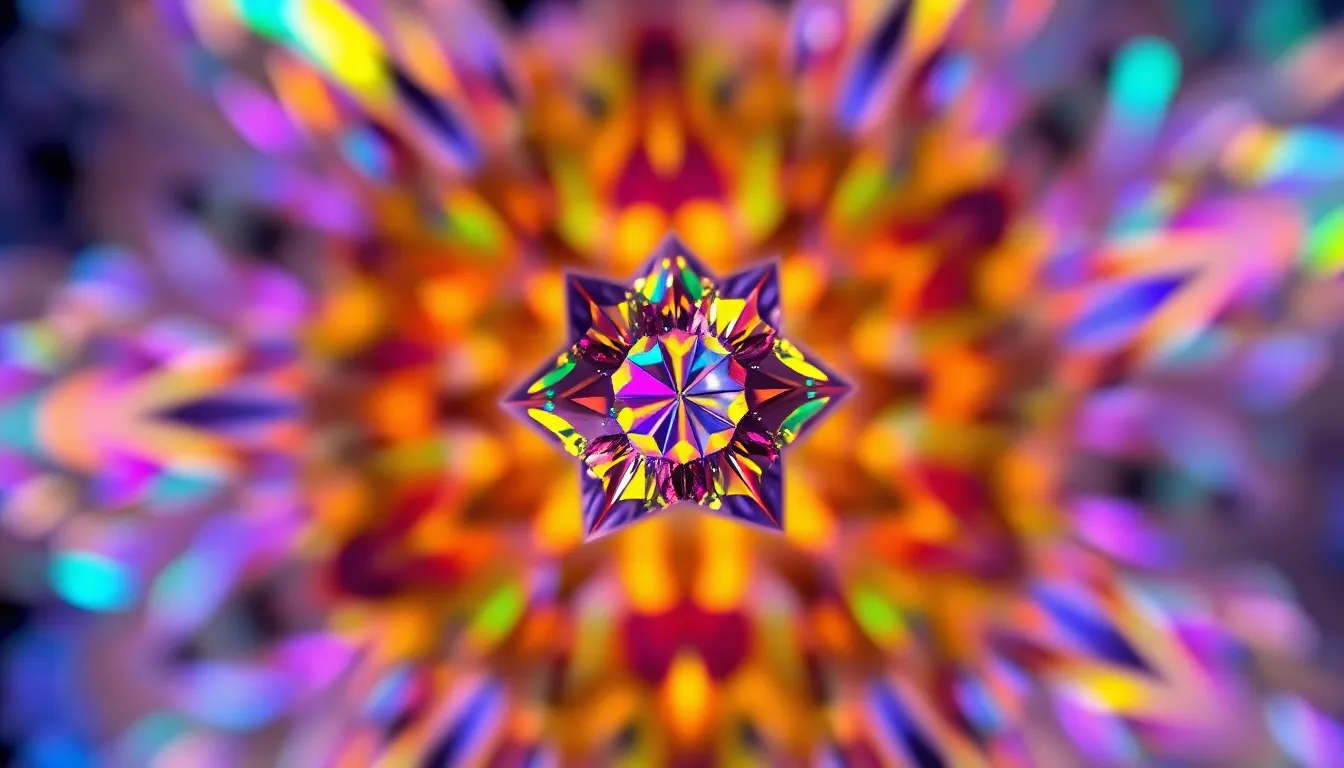
dramatic image of a crystal structure viewed under a polarized light microscope, captured with Leica Q3, 28mm f/1.7 Summilux. The lighting is achieved using cross-polarization, creating brilliant colors and sharp contrasts in the crystal. The depth of field is shallow at f/1.7, allowing the intricate details of the crystal to stand out sharply against the soft bokeh background. The color palette is vibrant, showcasing a range of saturated blues, greens, and yellows due to the polarization effect. The composition utilizes a centered approach, allowing the mesmerizing fractal patterns within the crystal to captivate the viewer.
16:9
close-up image of bacteria colonies on an agar plate, shot with Fujifilm GFX 100S, 110mm f/2 macro lens. The lighting is a combination of bright, focused LED lights that create sharp reflections off the gelatinous surface. A shallow depth of field at f/2 isolates the colonies, with a creamy bokeh highlighting the texture of the agar. The color grading is naturally muted with desaturated earth tones emphasizing the biological aspect. The composition highlights the leading lines of bacterial growth radiating from a central point, creating a dynamic visual flow.
16:9
detailed image of a stained tissue sample under a microscope, captured with Hasselblad X2D 100C, 90mm f/2.5 medium format. The scene is lit with soft, diffused daylight, emphasizing the rich textures within the sample. A hyperfocal depth of field ensures everything from the foreground to the background remains sharp, allowing for an expansive view. The color science adopts a Kodak Portra 400 palette, producing warm skin tones and creamy highlights, enhancing the tissue structures' visual appeal. The composition is centered, with a symmetrical arrangement of tissue features that draws the eye into the microscopic world.
16:9
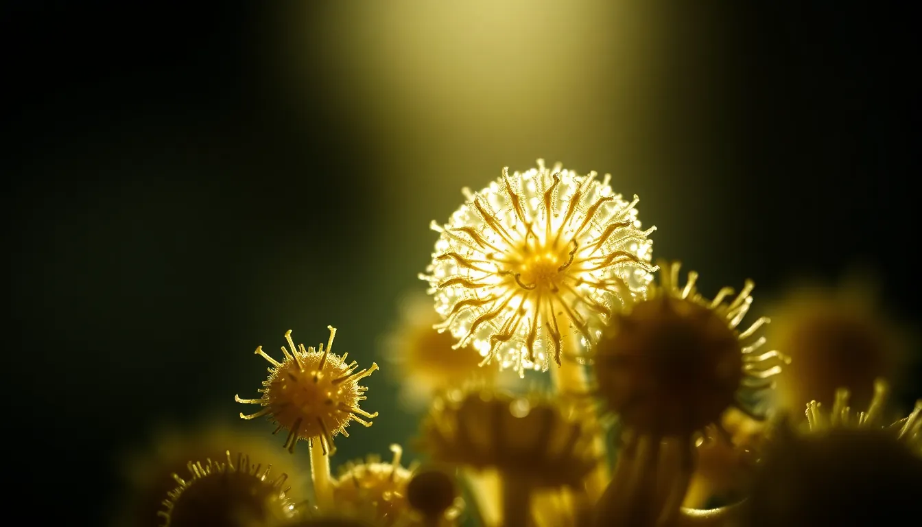
highly detailed image of pollen grains on a microscope slide, shot on Nikon Z9, 70-200mm f/2.8 telephoto at 135mm with intense backlighting creating a dramatic silhouette effect. The lighting accentuates the delicate structures of the pollen grains, which are rendered sharply in focus while the background fades into a soft, painterly blur. The color palette features natural muted tones, with earthy greens and browns giving a sense of organic texture. The composition employs the rule of thirds, positioning the largest pollen grain at a power point, surrounded by smaller grains, enhancing the visual interest.
16:9
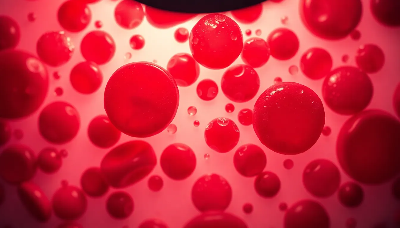
hyper-detailed photorealistic image of a vibrant, stained slide of human blood cells under a microscope, shot on Canon EOS R5, 24-70mm f/2.8L at 50mm. The lighting features a soft, diffused illumination from an overhead LED microscope light, highlighting the intricate details of the cells. A shallow depth of field at f/2.8 creates a creamy bokeh, allowing the red and white blood cells to pop against a soft-focus background. The image utilizes a Fujifilm Velvia-inspired color palette, emphasizing deep reds and soft whites for dramatic contrast. The composition centers on the cells with a slight diagonal leading line created by the edge of the slide, showcasing texture details such as the smooth glass slide and the cellular structures within.
16:9
magnified view of fungal spores captured using Leica Q3, 28mm f/1.7 Summilux. Natural overcast daylight provides soft, even illumination highlighting the spore structures without harsh shadows. Hyperfocal distance allows a tack-sharp focus across the entire frame, showcasing the details and textures of the spores. The color science leans towards natural muted tones to reflect the subtleties of the fungal life, with earthy browns and greens. Centered symmetrical composition draws attention to the unique patterns of the spores, enhancing their intricate designs.
16:9
an elegant close-up of a single pollen grain viewed through a microscope, captured with Fujifilm GFX 100S, 110mm f/2 macro. Soft natural daylight from a window creates gentle highlights and shadows that showcase the grain's intricate textures. Shallow depth of field at f/2 allows the background to melt into soft bokeh, providing a dreamy backdrop. The Kodak Portra 400 color palette enhances warm tones, making the pollen grain pop against a blurred green background. Rule of thirds composition places the grain at the left power point for dynamic interest.
16:9
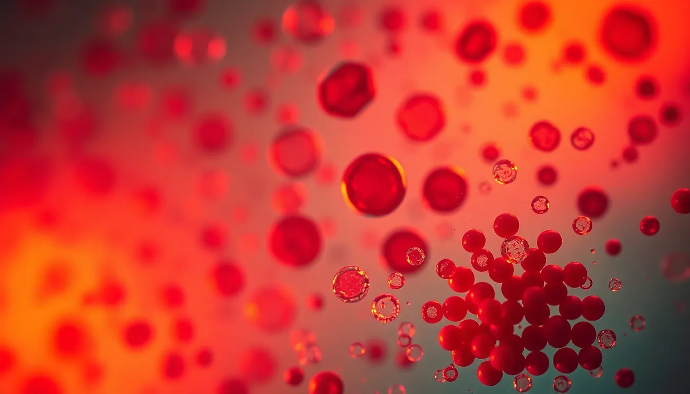
dramatic view of a blood smear under a microscope using Hasselblad X2D 100C, 90mm f/2.5 medium format. Warm tungsten light creates a rich, golden hue while illuminating blood cells, enhancing the contrast between red and white blood cells. Selective focus on the red blood cells at f/2.5 creates a beautiful bokeh effect in the background. The cinematic teal and orange color grading adds a striking visual appeal, while the rule of thirds composition emphasizes the clustering of cells in the lower right quadrant.
16:9
highly magnified image of bacterial cultures captured with Canon EOS R5, 24-70mm f/2.8L at 50mm. Studio lighting using a softbox creates dramatic highlights and shadows, enhancing the textures of the agar plate. Hyperfocal distance allows crisp focus from foreground to background, showcasing clusters of bacteria in various shapes and colors across the dish. A natural muted tone color palette with subtle earthy tones reflects the organic nature of the sample, while leading lines of the petri dish guide the viewer's eye inward. Textured agar surface reveals small imperfections, adding realism.
16:9
intricate close-up of a plant cell under a microscope shot on Nikon Z9, 70-200mm f/2.8 telephoto at 150mm. Captured using natural diffused daylight from overhead lighting, creating a soft, even illumination across the image. Shallow depth of field at f/2.8 highlights the cell structure while blurring the background into soft green hues. Vibrant Fujifilm Velvia-inspired colors render deep greens and bright cytoplasm, with the cell wall clearly defined. Centered symmetrical composition draws attention to the detailed microstructure, showcasing smooth cell membranes and intricate organelles like chloroplasts coated in a fine moisture.
16:9
captured with Leica Q3, 28mm f/1.7 Summilux, utilizing dappled sunlight filtering through leaves for soft lighting, shallow depth of field at f/1.7 creating a magical bokeh effect in the background, natural muted tones with desaturated earth colors highlighting the subject, composition featuring rule of thirds with a focus on a detailed leaf surface showcasing trichomes and veins under the microscope.
16:9
shot on Hasselblad X2D 100C, 90mm f/2.5 medium format, featuring dramatic side lighting from a single overhead lamp creating stark contrasts, selective focus on a sample of human hair revealing the fine texture and details, Fujifilm Velvia-inspired saturated colors enhancing the natural hues of the hair, composition framed using negative space for dramatic effect, allowing the detailed strands to stand out.
16:9
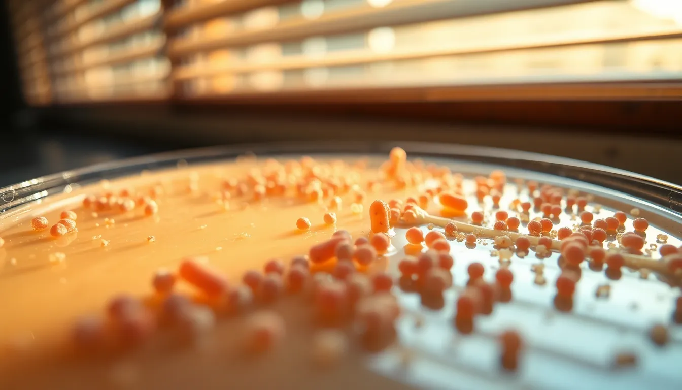
Fujifilm GFX 100S, 110mm f/2 macro capturing a close-up image of bacteria colonies growing on agar, natural soft daylight filtering through blinds creates an ethereal lighting effect, shallow depth of field at f/2 emphasizing the core details of the bacterial growth, natural muted tones with desaturated earth colors enhancing the organic feel, composition utilizing leading lines of the agar dish leading to the vibrant colonies.
16:9
captured with Canon EOS R5, 24-70mm f/2.8L at 50mm, under a softbox lighting setup providing even illumination with a gentle glow, hyperfocal distance at f/8 ensuring sharpness from foreground to background, Kodak Portra 400 color palette enhancing the warm skin tones and lifting shadows, centered symmetrical composition focusing on a detailed cross-section of a plant cell, showcasing cell structures and chloroplasts with visible texture.
16:9
shot on Nikon Z9, 70-200mm f/2.8 telephoto at 135mm, illuminated with bright white LED ring light creating sharp reflections, shallow depth of field at f/2.8 blurring the background into soft colors, natural muted tones with desaturated earth colors accentuating the specimen, rule of thirds composition with a focus on the intricate details of a butterfly wing sample, showcasing the delicate scales and variations in color.
16:9
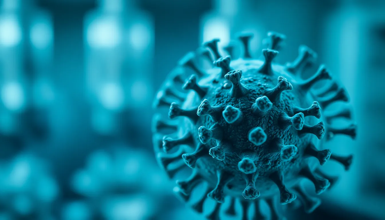
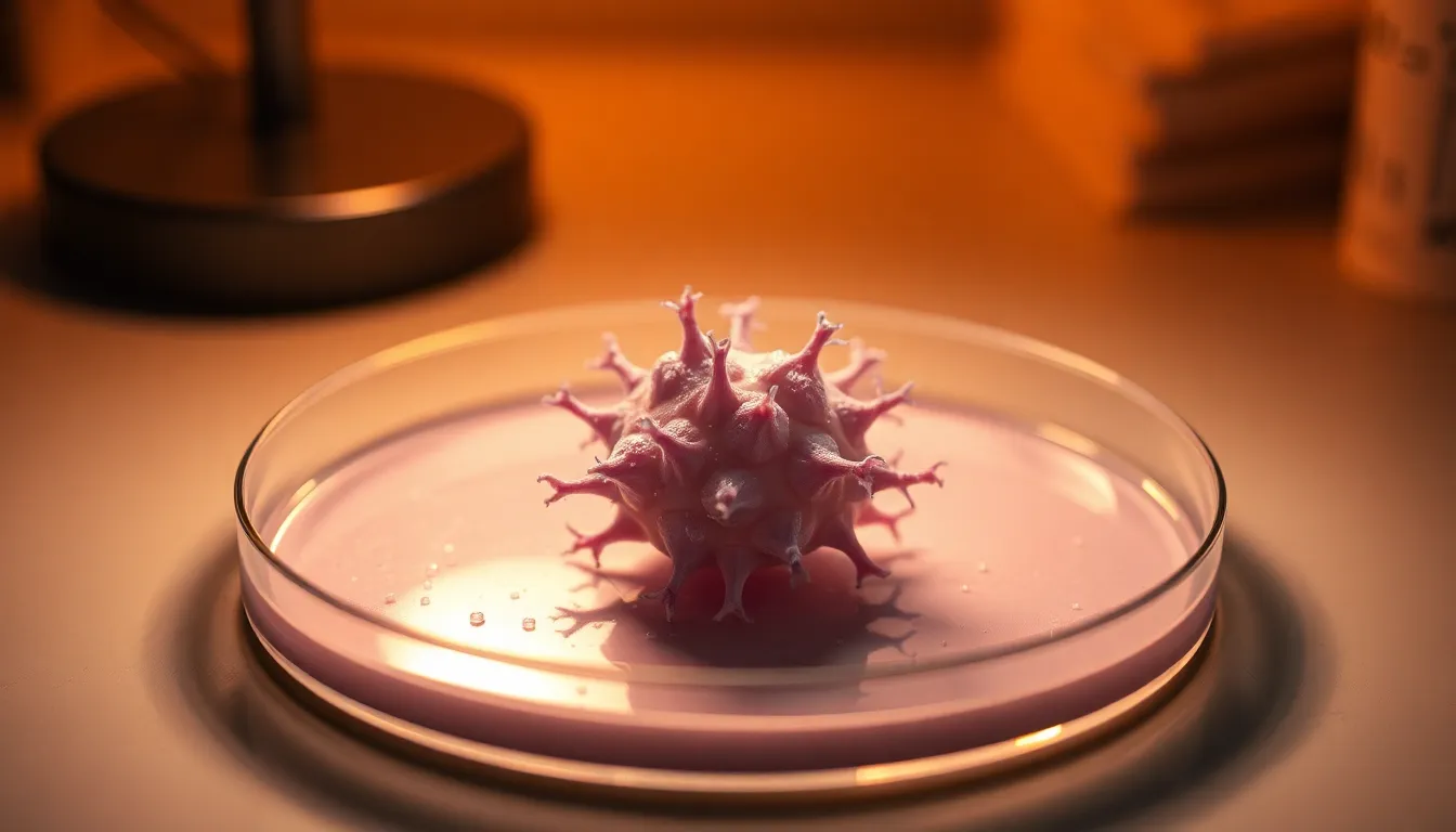
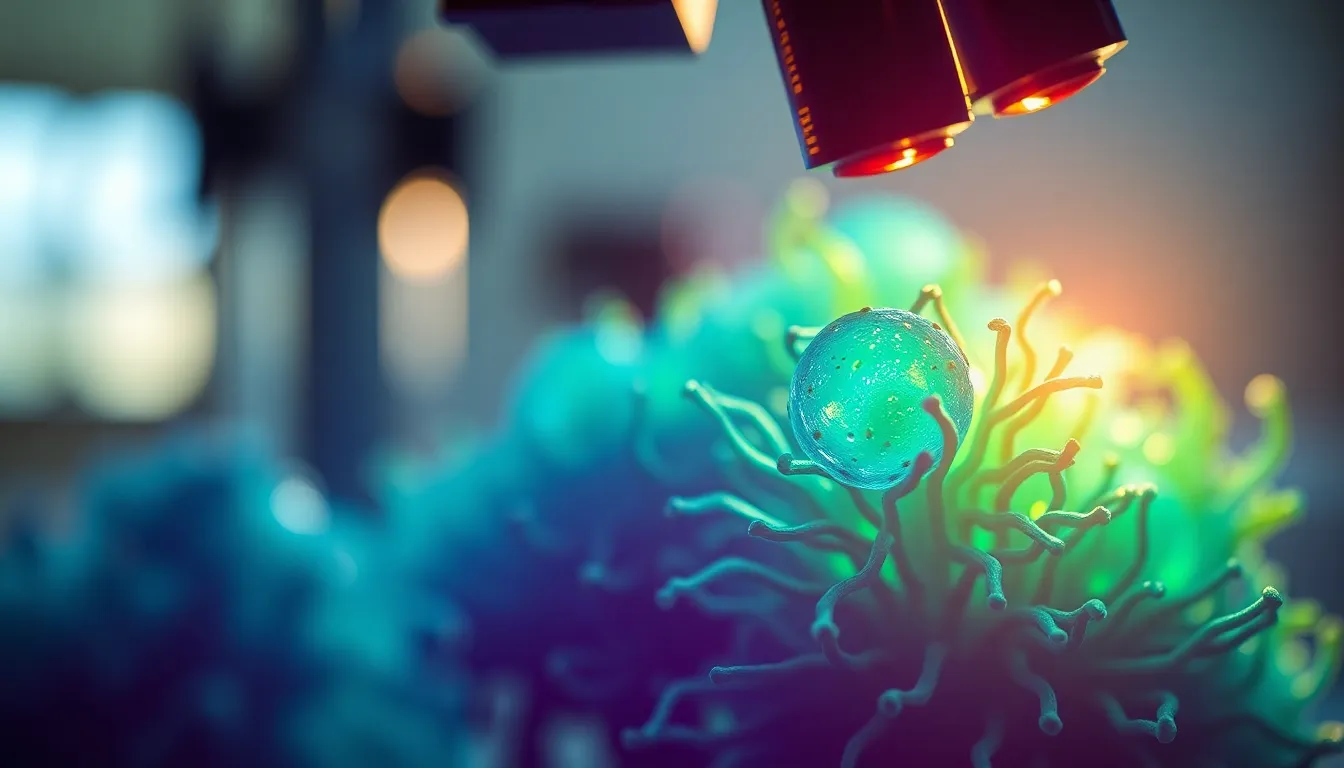
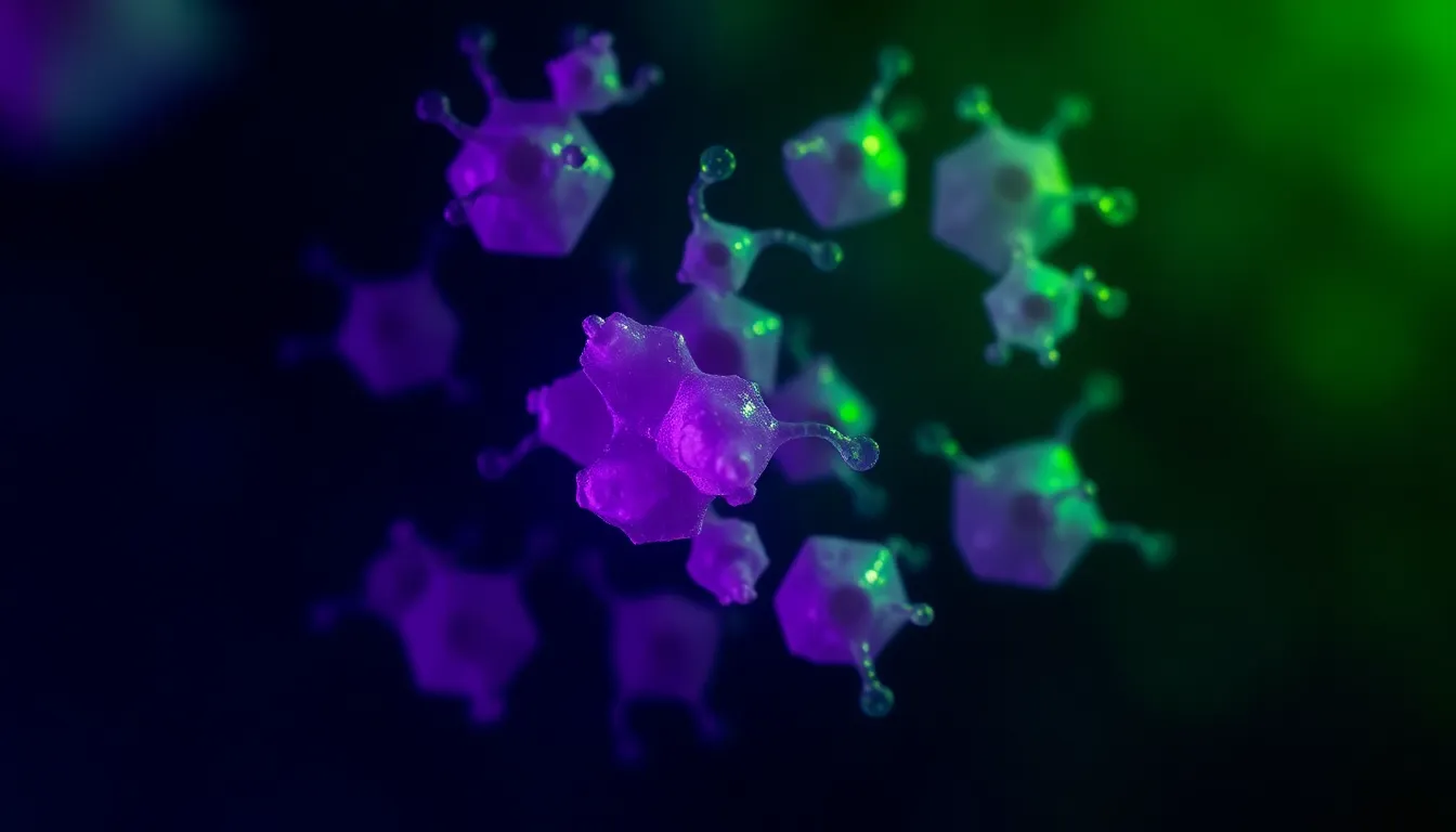
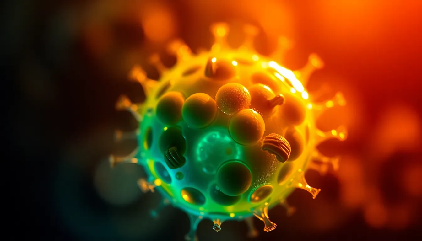
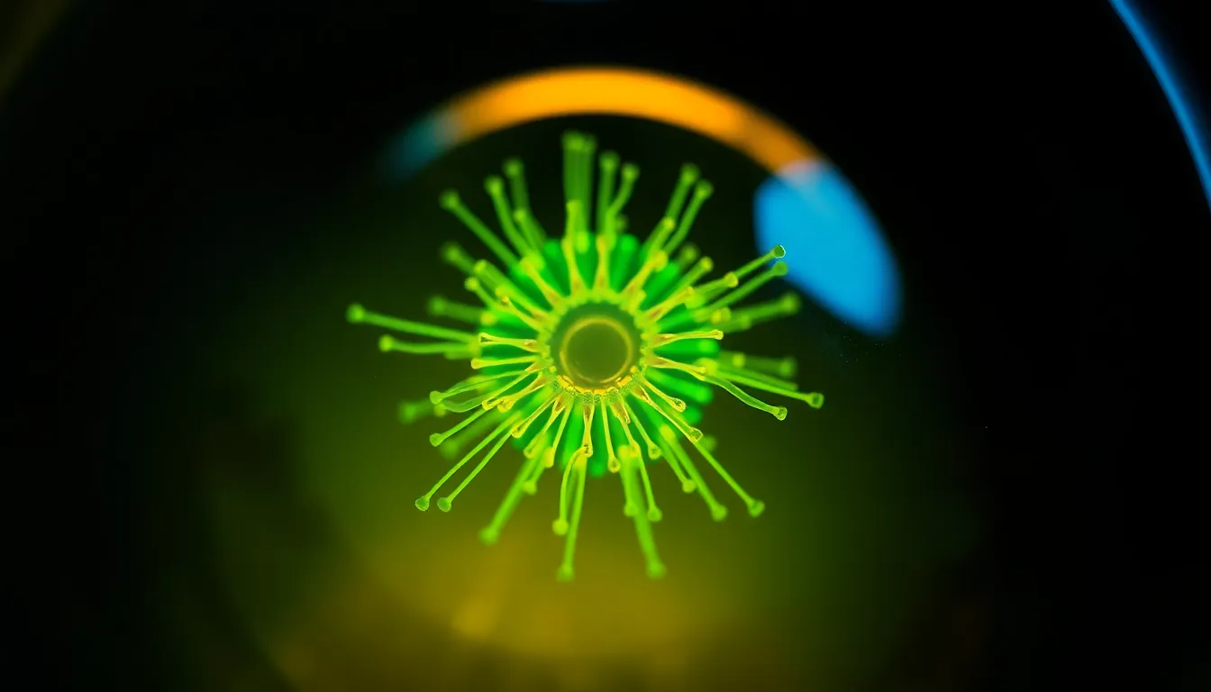
a detailed capture of a virus particle viewed under high magnification, shot on Leica Q3 with a 28mm f/1.7 Summilux lens, illuminated by harsh laboratory lights creating stark highlights and shadows, shallow depth of field at f/2 isolating the virus in crisp detail against a blurred background, natural muted tones with desaturated blues and greens showcasing the virus's surface textures, composition emphasizing the virus particle in the foreground with a stark contrast against the lab's sterile environment.
16:9

a photorealistic examination of a crystal structure under polarized light, captured with Fujifilm GFX 100S, 110mm f/2 macro lens, with dramatic lighting produced by polarized filters to create striking contrasts in colors, shallow depth of field at f/4 focusing on the central crystal while the surrounding areas dissolve into soft shapes, cinematic teal and orange color grading enhancing the vibrancy of the crystals, composition employs a dynamic angle to reveal the geometric shapes and symmetrical patterns of the crystals.
16:9
an artistic representation of a leaf cross-section viewed under a microscope, shot on Hasselblad X2D 100C with a 90mm f/2.5 medium format lens, backlit by a warm tungsten lamp enhancing the rich textures of the leaf, shallow depth of field at f/2.8 creating a dreamy bokeh background, with a Kodak Portra 400 color palette emphasizing warm greens and soft browns, composition showcasing the leaf's intricate vein patterns at the intersection of light and shadow.
16:9
a detailed image of bacteria colonies on an agar plate, captured with Canon EOS R5, 24-70mm f/2.8L at 35mm, natural diffused daylight filtering through a window providing soft, even lighting, hyperfocal distance at f/8 ensuring everything from the foreground to the background is sharp, with a natural muted color palette highlighting the subtle yellows and greens of the bacteria, composition using leading lines of the agar plate's edges directing the viewer's eye to the central growths, along with the texture of the agar surface.
16:9
an intricate close-up of a cell sample viewed under a microscope, shot on Nikon Z9 with 70-200mm f/2.8 telephoto at 135mm, illuminated by a focused LED ring light creating sharp highlights, shallow depth of field at f/2.8 bringing the cell structures crisply into focus while softening the background, vibrant colors inspired by Fujifilm Velvia with rich purples and greens, composition featuring a centered arrangement of the cells against a contrasting dark background, revealing the fine textures of the cell membranes and organelles.
16:9

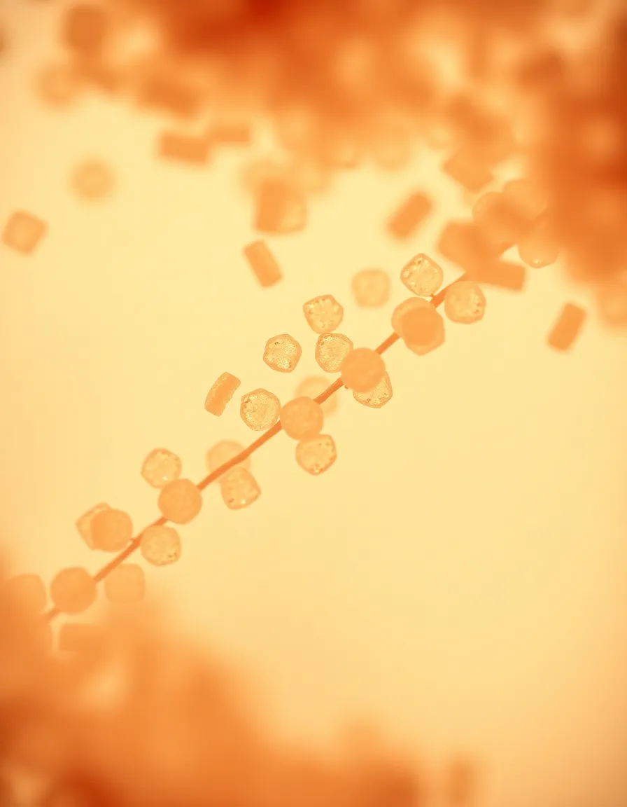
A detailed view of textile fibers under a microscope, captured with a Leica Q3, 28mm f/1.7 Summilux. Overcast daylight creates a soft, even light that highlights the unique textures of the fibers. The image features a hyperfocal distance, ensuring sharp detail from the foreground to infinity. The color palette showcases natural muted tones with desaturated earth colors, enhancing the organic appearance of the fibers. Compositionally, a rule of thirds placement allows for visual balance and intrigue.
2:3
An artistic rendering of pollen grains viewed through a microscope, taken with a Hasselblad X2D 100C, 90mm f/2.5 medium format. Ambient ambient light filters through a soft diffusion panel, creating soft highlights on the grains. Selective focus captures the detail in the foreground while allowing the background to dissolve into a soft blur. The color palette features warm skin tones and lifted shadows, providing a natural yet artistic quality. The composition utilizes a diagonal layout, creating a dynamic interaction between the grains.
3:4
Intricate image of a crystal structure under a microscope, captured with a Fujifilm GFX 100S, 110mm f/2 macro. Neon backlighting accentuates the sharp edges and facets of the crystal, creating an ethereal glow. The shallow depth of field at f/2 isolates the crystal from the background, producing a painterly bokeh effect. Color grading features cinematic teal and orange tones, providing a modern aesthetic. Compositionally, the crystal is placed according to the rule of thirds, enhancing visual interest and dynamism.
4:3
Close-up of a bacterial culture on an agar plate, photographed on a Canon EOS R5, 24-70mm f/2.8L at 50mm. Soft, diffused daylight through large windows creates natural lighting that captures the colors and textures of the cultures. Hyperfocal distance ensures everything is in sharp focus, revealing the intricate patterns of bacterial growth. The image features a natural muted color palette with desaturated earth tones, enhancing the organic feel. Compositionally, leading lines from the culture edges draw attention toward the center of the plate.
3:2
Microscopic view of a single cell, captured with a Nikon Z9, 70-200mm f/2.8 telephoto at 200mm. Dramatic side lighting enhances cell organelles, showcasing texture and detail. Shallow depth of field at f/2.8 blurs the background softly, while the subject remains in sharp focus. Color grading reminiscent of Fujifilm Velvia, emphasizing rich greens and vibrant blues. Composition utilizes centered symmetry, allowing viewers to appreciate the intricate patterns within the cell membrane. The surface shows fine details of the cellular structure and membrane layers.
16:9
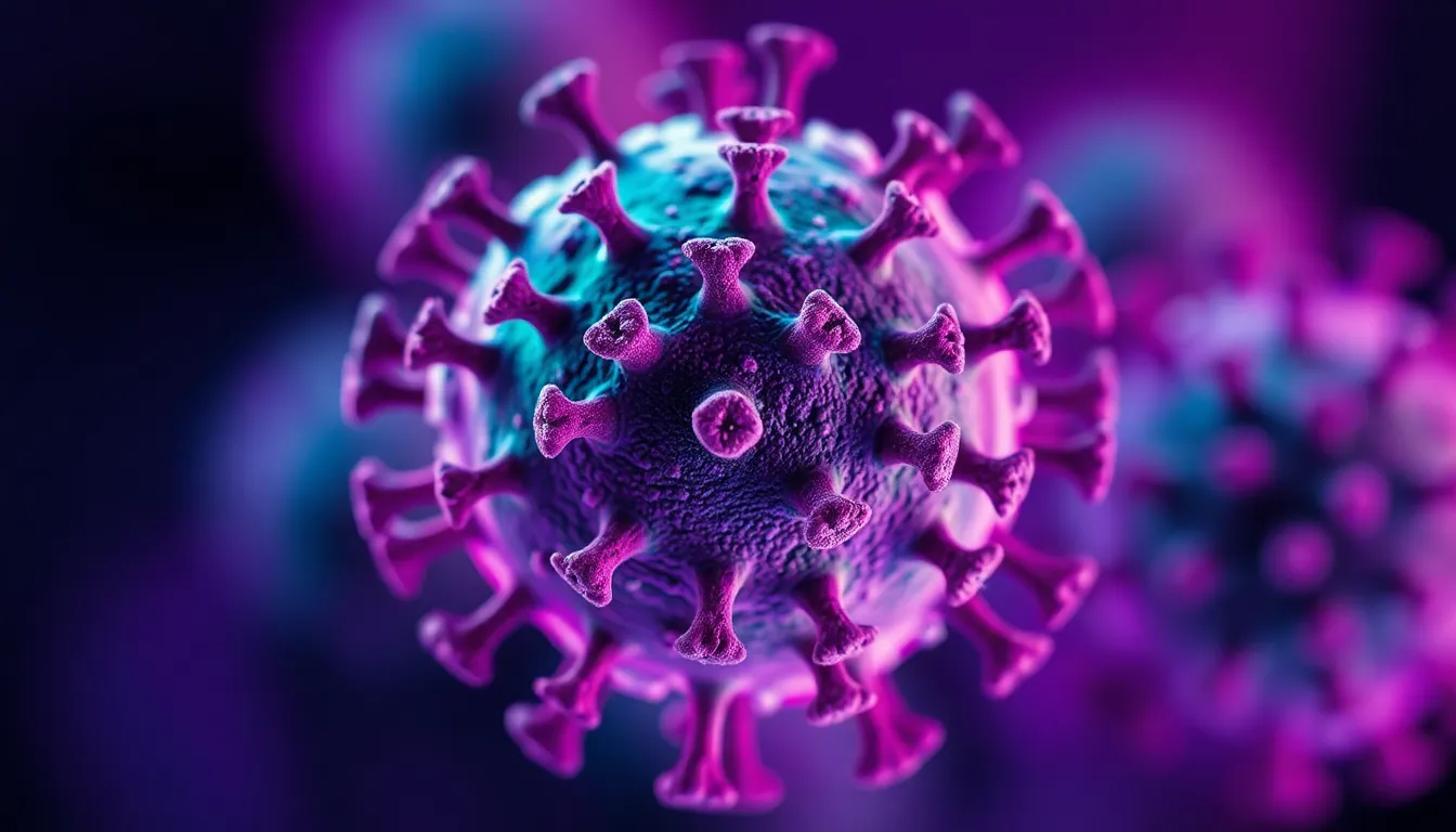
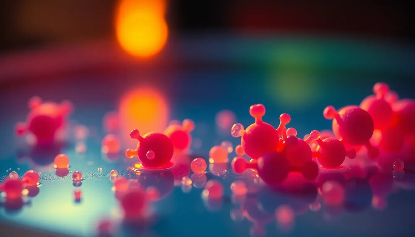
close-up of a viral structure, captured with Leica Q3, 28mm f/1.7 Summilux, showcasing soft diffused studio lighting to highlight textures, shallow depth of field at f/1.7 for enhanced focus on the virus. The color palette is dominated by vibrant purples and greens, evoking an otherworldly feel. The composition features a centered layout, drawing the viewer's eye directly to the intricate details of the viral particles, while the smooth textures of the background create a stark contrast to the subject's complexity.
16:9
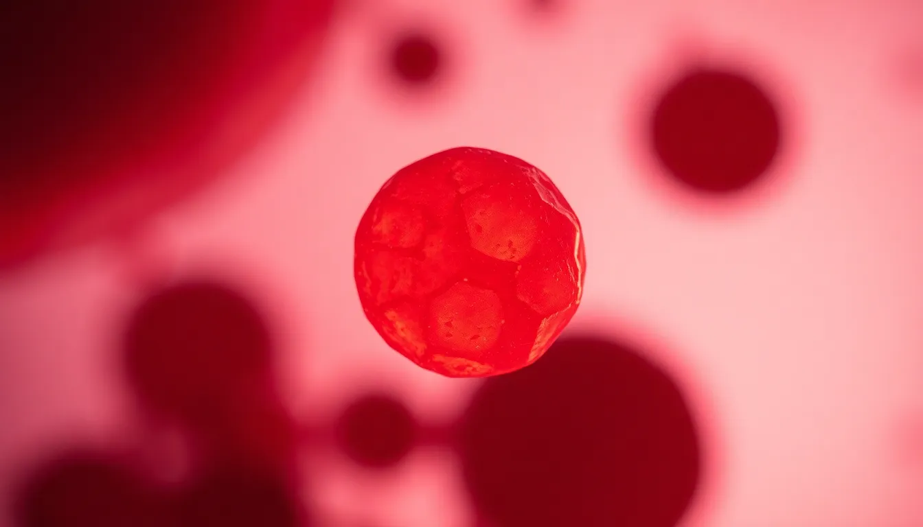
dynamic view of a human red blood cell under a microscope, captured with Hasselblad X2D 100C, 90mm f/2.5 medium format, using practical lighting from a tungsten lamp to create warm highlights, shallow depth of field at f/2.8 to emphasize the cell while softening the background. The color palette is rich in deep reds and subtle yellows, creating a vivid representation of the blood cell. The composition is centered, ensuring the blood cell remains the focal point against a softly blurred backdrop.
16:9

intricate image of crystalline structures, shot on Fujifilm GFX 100S, 110mm f/2 macro, using dramatic, side lighting to accentuate the facets and textures, shallow depth of field at f/2 creating a striking focus on the crystals. The color palette features cool blues and whites, highlighting the icy clarity of the crystals. The composition uses leading lines to draw the viewer's eye along the edges of the crystals, while the delicate textures of the crystalline surfaces are rendered in sharp detail.
16:9
detailed view of bacteria culture on an agar plate, captured with Canon EOS R5, 24-70mm f/2.8L at 50mm, illuminated by bright, diffused studio lighting to enhance colors, hyperfocal distance at f/8 ensuring all elements are sharp. The color palette features vibrant yellows, greens, and reds from bacterial colonies. The composition follows the rule of thirds, with the agar plate placed strategically off-center. Texture is highlighted through the glossy surface of the agar and the fuzzy edges of the bacteria, creating a striking visual.
16:9
microscopic view of a cross-section of a leaf, shot on Nikon Z9, 70-200mm f/2.8 telephoto at 135mm, illuminated by soft overcast natural light, shallow depth of field at f/2.8 creating a creamy bokeh background. The color palette features rich greens and earthy browns, showcasing the intricate vein structures. The composition is centered, emphasizing the details of the leaf while softly blurring the surrounding environment. Textures are vivid, capturing the fine cellular structure and surface details of the leaf's epidermis.
16:9
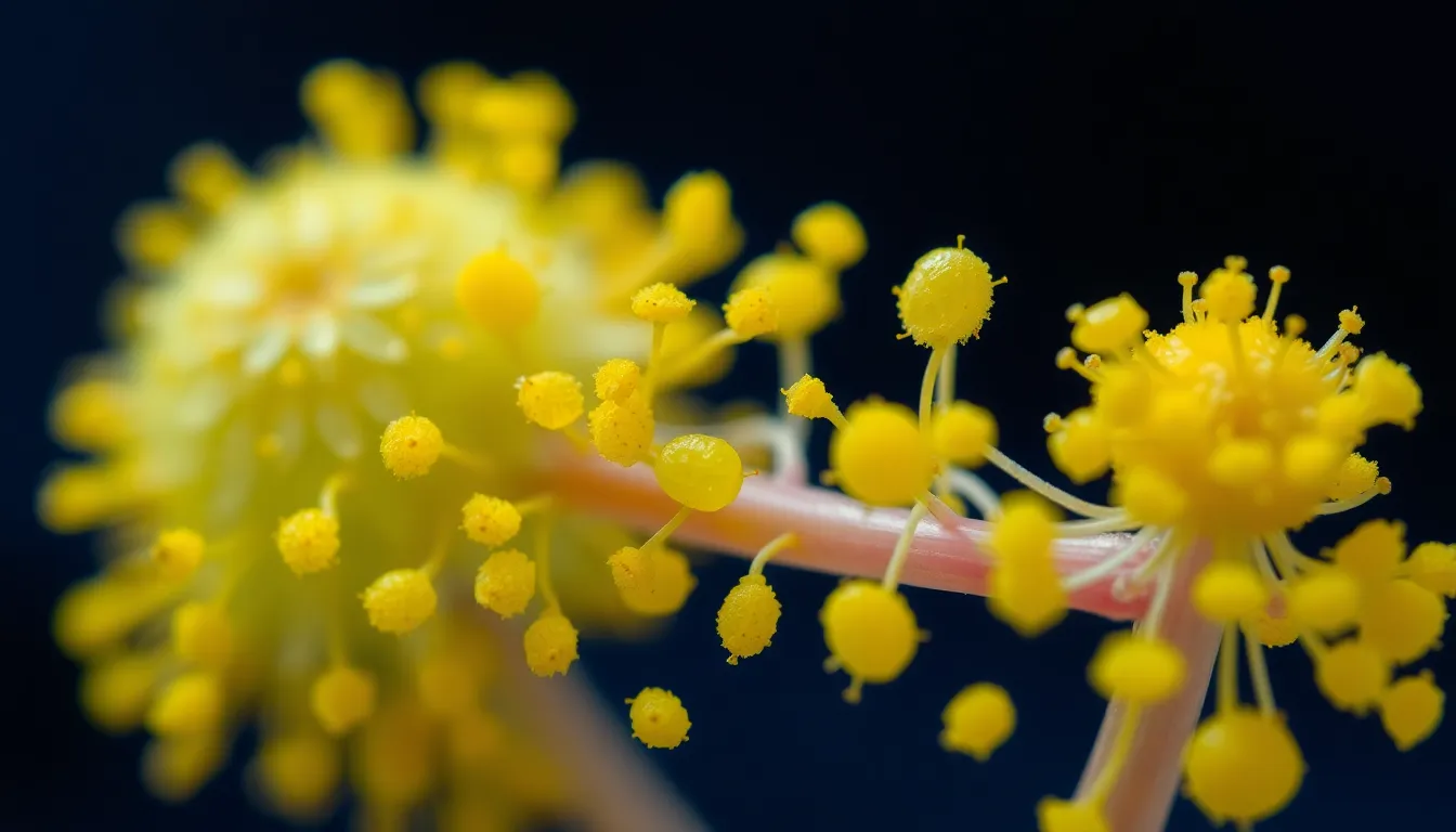
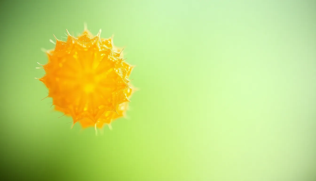
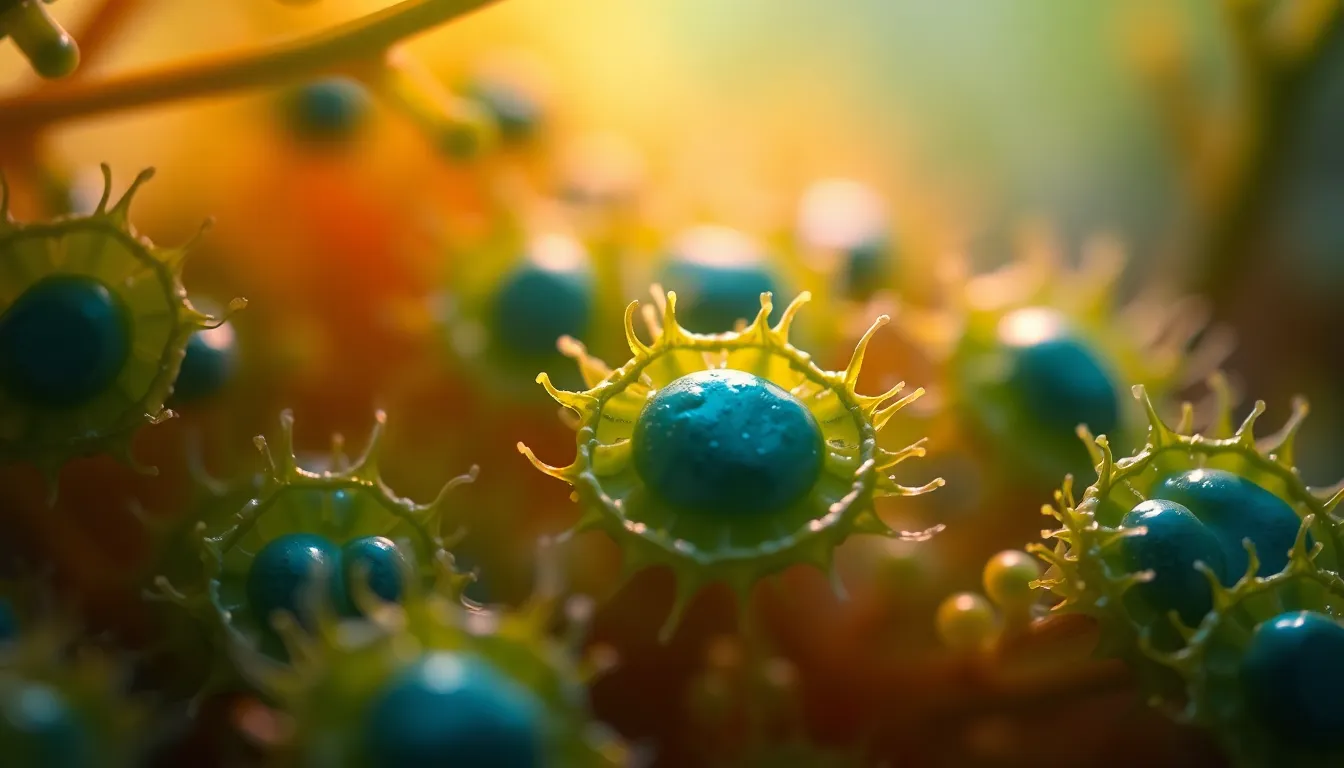
An extraordinary microscopic image of pollen grains from a flowering plant, captured with a Leica Q3, 28mm f/1.7 Summilux lens. The lighting is achieved using a ring light for even illumination, enhancing the colors and details of the pollen. The depth of field is shallow at f/2, allowing the viewer to appreciate the details while softly blurring the background. The color palette features vibrant yellows and greens, reflecting the natural hues of flower pollen. The composition employs a rule of thirds arrangement, prominently featuring the pollen grains against a contrasting dark blue background that emphasizes their vibrant colors and textures.
16:9

A detailed microscopic representation of crystalline structures in salt, captured with a Hasselblad X2D 100C, 90mm f/2.5 medium format lens. The lighting uses side lighting to accentuate the sharp edges of the crystals and create striking highlights. Depth of field is set to f/8, providing a tack-sharp image from foreground to background. The color palette consists of clear whites and soft blues, reflecting the natural colors of salt crystals. The composition is centered, showcasing the geometric patterns of the crystals against a dark slate background, enhancing their brilliance. The texture highlights the sharp, faceted surfaces of the crystals.
16:9
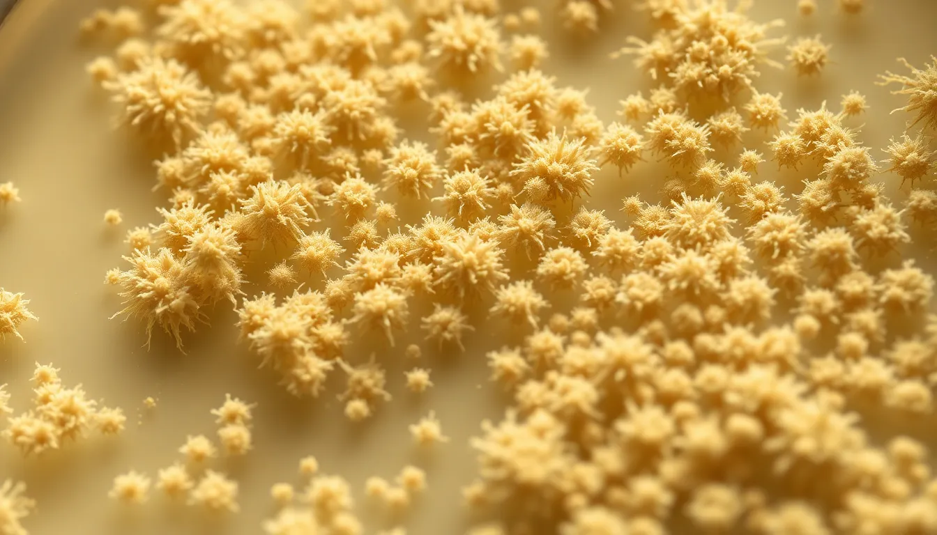
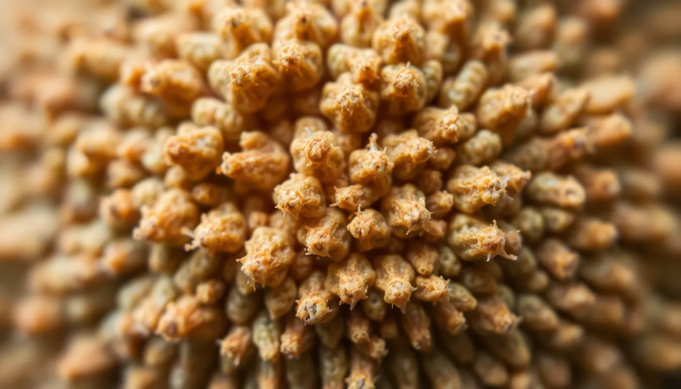
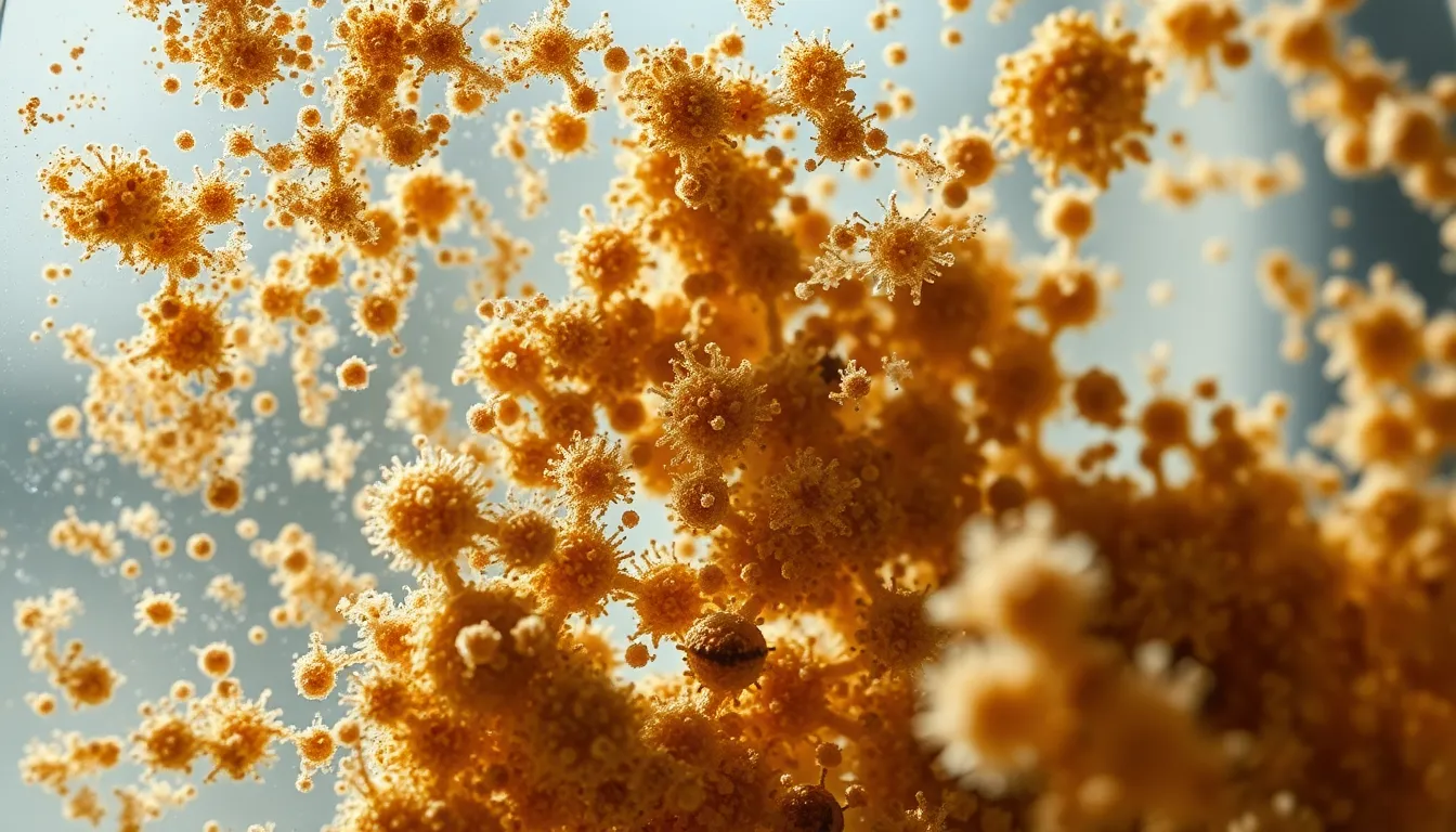
An intricate microscopic view of mold spores on a petri dish, captured with a Fujifilm GFX 100S, 110mm f/2 macro lens. The lighting is soft and diffused, mimicking natural daylight, while depth of field is shallow at f/2.8, bringing the spores into sharp focus against a blurred background. The colors are natural and muted, showcasing earth tones of beige and green. The composition employs leading lines, drawing the eye along the patterns formed by the spores, enhancing the organic feel. The surface texture of the mold is visible, revealing fine details and fuzziness.
16:9
A stunning micrograph of a butterfly wing, showcasing the intricate scales and colors, captured with a Canon EOS R5, 24-70mm f/2.8L at 50mm. The lighting is achieved through a focused spotlight, creating dramatic shadows and highlighting the vibrant iridescence of the scales. Depth of field is set to f/4, keeping the entire wing in crisp focus. The color palette features bright blues and greens with hints of orange, contributing to an enchanting visual. The composition follows the rule of thirds, with an emphasis on the detailed patterns of the scales against a soft gray background.
16:9

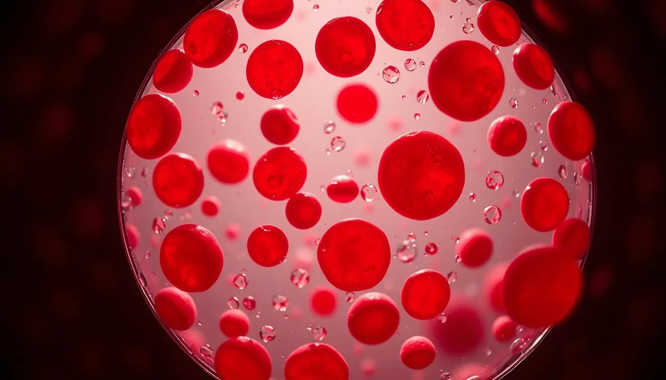
A vibrant and colorful microscopic view of human blood cells, captured with a Nikon Z9, 70-200mm f/2.8 telephoto at 135mm. The lighting features a soft, diffused backlight illuminating the cells with subtle highlights, while the depth of field is shallow at f/2.8, creating a creamy bokeh effect in the background. The color palette consists of rich reds and soft pinks with a slight bluish tint. The composition uses a centered arrangement, highlighting the intricate details of the cells against a dark background. The texture shows the natural translucency and surface details of the blood cells, emphasizing their unique shapes.
16:9
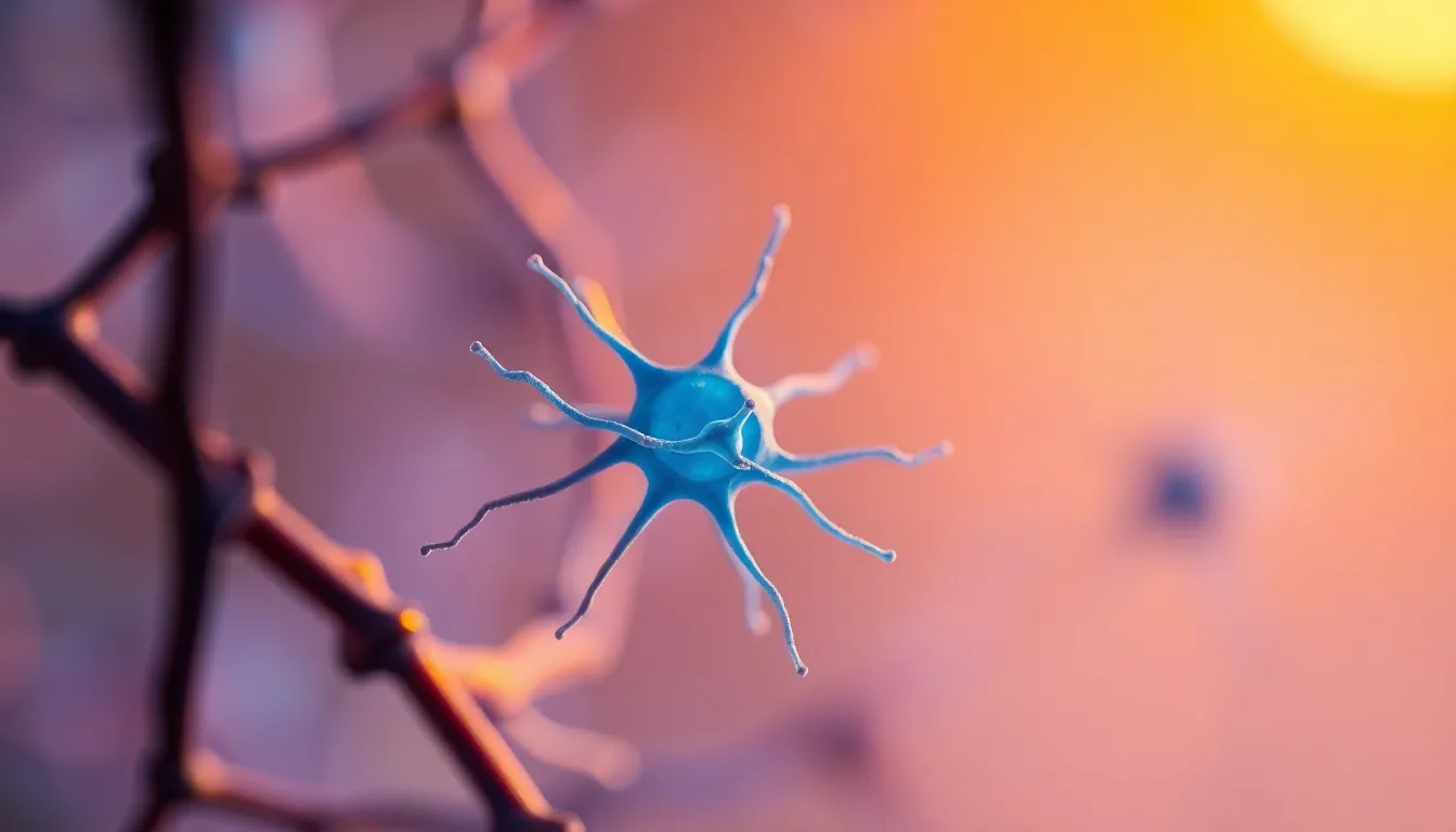
A captivating image of a single neuron under the microscope, captured with Leica Q3, 28mm f/1.7 Summilux lens. The lighting is a combination of natural sunlight and a warm tungsten desk lamp, creating a unique blend of colors and shadows. The depth of field is shallow at f/1.7, resulting in a soft, creamy background that emphasizes the neuron in exquisite detail. The color palette features cool blues with warm undertones, creating visual contrast. The composition follows a centered layout, drawing attention to the elegance of the neuron's structure.
16:9
A detailed view of mold spores captured on a glass slide, photographed using Hasselblad X2D 100C, 90mm f/2.5 medium format lens. The setup features natural lighting with bright ambient daylight filtering through a window, resulting in crisp, clear textures. A hyperfocal distance setting at f/8 ensures clarity across the entire frame, showcasing the various sizes and shapes of the spores. The color palette consists of earthy browns and muted yellows, which convey a sense of organic decay. The rule of thirds composition places the most prominent spores at the left power point, enhancing visual interest.
16:9
An artistic view of plant cells under a microscope, captured using Fujifilm GFX 100S, 110mm f/2 macro lens. The scene is illuminated with a softbox providing gentle Rembrandt lighting, creating depth through shadows. The shallow depth of field at f/2 highlights the detailed cell walls and chloroplasts against a soft, blurred background. This image employs a Fujifilm Velvia-inspired color palette with saturated greens and blues. The composition utilizes leading lines to draw the eye through the image, reflecting the structured nature of plant life.
16:9
A dynamic image of human blood cells under a microscope, shot on Nikon Z9, 70-200mm f/2.8 telephoto at 100mm. The scene is lit with a diffused daylight setup, allowing natural colors to emerge vividly. Utilizing hyperfocal distance at f/8, the detail of the red blood cells and the textures of the glass slide are all in sharp focus. The image features a natural muted color palette with soft reds and earthy tones. A centered symmetrical composition draws direct attention to the blood cells, portraying their biological significance.
16:9
A close-up of a vibrant diatom viewed under a microscope, captured with Canon EOS R5, 24-70mm f/2.8L at 50mm. The illumination is achieved with a bright LED ring light, creating sharp contrast on the intricate details of the silica structure. A shallow depth of field at f/2.8 emphasizes the subject, allowing the surrounding glass slide to melt into a soft blur. The color palette features rich greens and yellows, reminiscent of natural flora. The composition follows the rule of thirds, placing the diatom slightly off-center, while the textured surface of the glass reflects subtle highlights.
16:9
shot on Leica Q3, 28mm f/1.7 Summilux, with Rembrandt lighting achieved using a single octabox positioned at 45 degrees, shallow depth of field at f/1.7 for a soft background, employing a cinematic teal and orange color grading to create a dramatic mood, the composition uses a centered symmetrical approach to highlight a detailed close-up of a crystal structure under a microscope, showcasing the intricate textures and facets of the crystal.
3:2
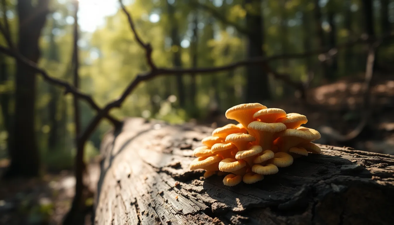
captured with Hasselblad X2D 100C, 90mm f/2.5 medium format, illuminated by soft natural light filtering through trees creating dappled patterns, depth of field at f/5.6 ensuring a sharp focus across the whole scene, using natural muted tones with desaturated earth colors to reflect a serene environment, composition encompasses leading lines of tree branches guiding the eye towards a vibrant microcosm of fungal structures on a decaying log, emphasizing the textures of the fungi and wood.
16:9
shot on Fujifilm GFX 100S, 110mm f/2 macro lens, illuminated with a warm tungsten desk lamp casting directional light, shallow depth of field at f/2 creating beautiful bokeh, using a Kodak Portra 400 color palette to enhance warm skin tones and soft highlights, the composition employs rule of thirds with a focus on a single specimen slide featuring a unique microorganism, showcasing the texture of the slide’s surface and the organism's fine details.
3:2
captured with Canon EOS R5, 24-70mm f/2.8L at 50mm, lit with soft diffused daylight from an overhead window, hyperfocal distance ensuring everything from the samples to the background remains tack-sharp at f/8, utilizing a Fujifilm Velvia-inspired saturated colors approach to highlight the vivid pigments, composition utilizing leading lines that draw the eye toward a range of colorful stained slides arranged on a table, with smooth glass surfaces reflecting light.
16:9
shot on Nikon Z9, 70-200mm f/2.8 telephoto at 100mm, illuminated with cold LED lighting for stark contrast, shallow depth of field at f/2.8 creating a soft bokeh that isolates the subject, using a natural muted tones color palette that emphasizes blues and greens, centered symmetrical composition highlighting a single cell structure with intricate details, surface textures like the roughness of cell membranes and fluidity of cytoplasm evident.
16:9